| Citation: |
LI Xiaofeng, LIU Caiyun, SHI Yan, LUO Xiaohu, WANG Gang, YAO Biao. Differential Diagnosis of Benign and Malignant Breast Lesions by Texture Feature Combined with Histogram Parameter Based on Tirm Sequence[J]. Cancer Research on Prevention and Treatment, 2018, 45(12): 1009-1013. DOI: 10.3971/j.issn.1000-8578.2018.18.0815

|
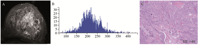
To explore histogram parameters and texture features of benign and malignant breast lesion on turbo inversion recovery magnitude(Tirm) sequence, and evaluate which parameter could help best differentiate benign from malignant breast lesion.
This retrospective study included 100 breast cancer patients who underwent conventional MRI and confirmed pathologically. Texture features were derived from the gray level co-occurrence matrix(GLCM), and entropy, energy, correlation, inertia, inverse difference moment, cluster prominence and mean value, skewness, kurtosis of histogram parameters were calculated. Then we assessed the diagnosis efficacy with these parameters among the variety kinds of benign and malignant breast lesions respectively, and establish the receiver-operating characteristic curve(ROC). We assessed the differences between benign and malignant groups by the Yoden index combined with clinic for the cut-off values.
The differences of correlation, cluster prominence, mean value and kurtosis were statistically significant between the benign and malignant groups(all P < 0.001); the differences of energy, entropy, inertia, inverse difference moment and skewness were not statistically significant(all P > 0.05). The results of ROC with correlation, cluster prominence, mean value and kurtosis on the diagnosis of benign and malignant breast lesions were statistically significant, respectively(P < 0.001). Combined the four parameters on diagnosis benign and malignant lesions, the AUC was 0.868, the sensitivity was 90.57% and the specificity was 72.34%.
The histogram analysis and texture analysis based on Tirm sequence could be used for the differential diagnosis of benign and malignant breast lesions. Correlation, cluster prominence, mean value and kurtosis have certain significance in the diagnosis. The combined diagnosis could improve the differential ability of benign and malignant breast lesions.
| [1] |
Gallego-Ortiz C, Martel AL. Using quantitative features extracted from T2-weighted MRI to improve breast MRI computer-aided diagnosis (CAD)[J]. PLoS One, 2017, 12(11): e0187501. doi: 10.1371/journal.pone.0187501
|
| [2] |
Imbriaco M, Cuocolo R. Does Texture Analysis of MR Images of Breast Tumors Help Predict Response to Treatment?[J]. Radiology, 2018, 286(2): 421-3. doi: 10.1148/radiol.2017172454
|
| [3] |
Wu J, Gong G, Cui Y, et al. Intratumor partitioning and texture analysis of dynamic contrast-enhanced (DCE)-MRI identifies relevant tumor subregions to predict pathological response of breast cancer to neoadjuvant chemotherapy[J]. J Magn Reson Imaging, 2016, 44(5): 1107-15. doi: 10.1002/jmri.25279
|
| [4] |
Pickles MD, Lowry M, Gibbs P. Pretreatment Prognostic Value of Dynamic Contrast-Enhanced Magnetic Resonance Imaging Vascular, Texture, Shape, and Size Parameters Compared With Traditional Survival Indicators Obtained From Locally Advanced Breast Cancer Patients[J]. Invest Radiol, 2016, 51(3): 177-85. doi: 10.1097/RLI.0000000000000222
|
| [5] |
Ko ES, Kim JH, Lim Y, et al. Assessment of Invasive Breast Cancer Heterogeneity Using Whole-Tumor Magnetic Resonance Imaging Texture Analysis[J]. Medicine(Baltimore), 2016, 95(3): e2453. http://www.wanfangdata.com.cn/details/detail.do?_type=perio&id=8c0b24339f353624ba381e15cd9f78fc
|
| [6] |
孙赛花, 周纯武, 赵莉芸, 等.动态增强磁共振成像纹理分析预测乳腺癌新辅助化疗疗效[J].中华肿瘤杂志, 2017, 39(5): 344-9. doi: 10.3760/cma.j.issn.0253-3766.2017.05.005
Sun SH, Zhou CW, Zhao LY, et al. Texture analysis based on contrast-enhanced MRI can predict treatment response to neoadjuvant chemotherapy of breast cancer [J]. Zhonghua Zhong Liu Za Zhi, 2017, 39(5): 344-9. doi: 10.3760/cma.j.issn.0253-3766.2017.05.005
|
| [7] |
Prevos R, Smidt ML, Tjan-Heijnen VC, et al. Pre-treatment differences and early response monitoring of neoadjuvant chemotherapy in breast cancer patients using magnetic resonance imaging: a systematic review[J]. Eur Radiol, 2012, 22(12): 2607-16. doi: 10.1007/s00330-012-2653-5
|
| [8] |
Waugh SA, Purdie CA, Jordan LB, et al. Magnetic resonance imaging texture analysis classification of primary breast cancer[J]. Eur Radiol, 2016, 26(2): 322-30. http://www.wanfangdata.com.cn/details/detail.do?_type=perio&id=0e918f60a991f47f32e564c7c67352a8
|
| [9] |
Salem A, O'Connor JPB. Assessment of Tumor Angiogenesis: Dynamic Contrast- enhanced MR Imaging and Beyond[J]. Magn Reson Imaging Clin N Am, 2016, 24(1): 45-56 doi: 10.1016/j.mric.2015.08.010
|
| [10] |
张竹伟, 华婷, 徐婷婷, 等.常规MRI纹理分析鉴别乳腺良、恶性病变的价值初探[J].中华放射学杂志, 2017, 51(8): 588-91. doi: 10.3760/cma.j.issn.1005-1201.2017.08.006
Zhang ZW, Hua T, Xu TT, et al. Differentiation of benign and malignant breast lesions using texture analysis of conventional MRI:a preliminary study[J]. Zhonghua Fang She Xue Za Zhi, 2017, 51(8): 588-91. doi: 10.3760/cma.j.issn.1005-1201.2017.08.006
|
| [11] |
冯红梅, 郭彩平, 徐志锋, 等.乳腺X线摄影和MRI直方图在鉴别乳腺纤维腺瘤和浸润性导管癌中的价值[J].医学影像学杂志, 2017, 27(1): 75-8. http://d.old.wanfangdata.com.cn/Periodical/yxyxxzz201701020
Feng HM, Guo CP, Xu ZF, et al. The value of X-ray photography and MRI histogram in distinguishing of breast fibroadenoma and infiltrating ductal car-cinoma[J]. Yi Xue Ying Xiang Xue Za Zhi, 2017, 27(1): 75-8. http://d.old.wanfangdata.com.cn/Periodical/yxyxxzz201701020
|

This work is licensed under a Creative Commons Attribution 3.0 License.
Copyright © Editorial Department of Cancer Prevention Research 鄂公网安备 42011102005013号 鄂ICP备2022015867号
Supported by: Beijing Renhe Information Technology Co., Ltd. 
 DownLoad: