文章信息
- 田金徽,李金龙,葛龙,王婧,张明霞,石芳毓,王小虎,张红. 2015
- TIAN Jinhui, LI Jinlong, GE Long, WANG Jing, ZHANG Mingxia, SHI Fangyu,WANG Xiaohu, ZHANG Hong. 2015
- PET/CT对宫颈癌淋巴结转移诊断价值的Meta分析
- Diagnostic Value of PET/CT for Metastatic Lymph Nodes in Cervical Cancer Patients: A Meta-analysis
- 肿瘤防治研究, 2015, 42(03): 270-276
- Cancer Research on Prevention and Treatment, 2015, 42(03): 270-276
- http://www.zlfzyj.com/CN/10.3971/j.issn.1000-8578.2015.01.013
-
文章历史
- 收稿日期:2014-02-16
- 修回日期:2014-11-14
2. 730000 兰州,中国科学院近代物理研究所;
3.甘肃省循证医学与临床转化重点实验室;
4. 730000 兰州,兰州大学基础医学院循证医学中心
5. 730000 兰州,兰州大学第一临床医学院;
6. 730000 兰州,兰州大学第二临床医学院
2.Institute of Modern Physics, Chinese Academy of Science, Lanzhou 730000, China;
3.Key Laboratory of Clinical Translational Research and Evidence-based Medicine of Gansu Province, Lanzhou 730000, China;
4.School of Basic Medical Science, Evidence-based Medicine Center of Lanzhou University, Lanzhou 730000,China;
5. The First Clinical Medical College of Lanzhou University, Lanzhou 730000, China;
6.The Second Clinical Medical College of Lanzhou University, Lanzhou 730000, China
宫颈癌为妇女最常见第二大肿瘤[1,2],据国际癌症研究所(IARC) 统计全球每年宫颈癌新发病例绝大多数在发展中国家[3],宫颈癌目前仍占中国女性生殖系统恶性肿瘤的第1位[4]。有报道称,Ⅰ期和Ⅱ期宫颈癌患者术后盆腔淋巴结转移率分别为0~16%和24.5%~31%,腹主动脉旁淋巴结转移率分别为0~22%和11%~19%,手术切除的淋巴结中,没有癌转移的逾90%[5]。所以,大部分早期宫颈癌患者并没有淋巴结转移,而系统淋巴结清扫术不仅增加了诊断的时间和成本,而且增加了患者手术并发症的风险。如果能够早期精确的探测淋巴结是否存在转移,对妇科恶性肿瘤宫颈癌进行准确分期、制定合理具体的治疗方案及评价患者的预后等,均有重要的临床价值。
PET因诊断淋巴结转移的准确性高而引起影像诊断医师和妇科肿瘤界专家的高度重视,其原理为被放射性同位素等正电子核素标记的人体基本代谢底物在病灶处积聚,并与病灶中的电子发生电子对湮灭,放出以180°角度向相反方向放射γ射线,根据射线的检出可以确定病灶内代谢情况,达到活体断层生化分析及细胞分子水平上的代谢与受体功能研究。常用的放射性标记药物为18FFDG(18F-氟代脱氧葡萄糖),它具有辐射小,衰变快的特点,是一种比较安全的检查剂。由于癌细胞代谢较正常组织细胞需要更多的营养,因此,能够对癌组织进行大致的定位和定性诊断。PET/CT凭借其分子显像技术的优势,兼顾了解剖学特点与功能学特点,在各种实质性肿瘤中的诊断价值得到证实,在宫颈癌领域,PET/CT已用于宫颈癌患者的初始分期,治疗计划的制定,治疗后的检测和随访以及预后的判断[5,6,7]。
为了客观评价PET/CT对宫颈癌淋巴结转移诊断价值,我们应用系统评价方法评价PET/CT诊断宫颈癌淋巴结转移文献的质量并进行合并分析,为临床提供参考。1 资料与方法1.1 纳入与排除标准1.1.1 纳入标准
(1)研究类型:关于PET/CT与金标准比较诊断宫颈癌淋巴结转移的试验,可提取或通过计算可获得四格表数据;(2)研究对象:伴有盆腔和(或)腹主动脉旁淋巴结转移的宫颈癌患者,无年龄、性别、种族及病因的限制;(3)诊断试验方法:PET/CT vs. 手术后病理结果(金标准)。1.1.2 排除标准
(1)动物实验等基础研究;(2)非中英文以外的文献;(3)非原始研究;(4)宫颈癌无淋巴结转移研究。1.2 文献检索
检索策略:参考《The Bayes Library of Diagnostic Studies and Reviews》[8],检索词分目标疾病、待评价试验、诊断准确性指标三部分,并根据具体数据库调整,所有检索采用主题词[MEDLINE(MeSH),EMATGE(EMTREE)]与自由词相结合的方式,所有检索策略通过多次预检索后确定。中文检索词:PET/CT、正电子发射计算机体层摄影、计算机断层显像、宫颈癌、宫颈肿瘤、淋巴结、敏感度、特异性、诊断等。英文检索词:positron emission tomography、PETCT、CT、cervical cancer、cervical neoplasm、lymph node、sensitivity、specificity、diagnosis等。检索资源:PubMed、Web of Science、The Cochrane Library、Embase.com、中文科技期刊数据库(维普)、万方全文数据库、中国期刊全文数据库、中国生物医学文献数据库、时间和语种不限。同时采用Google引擎搜索,并追踪纳入文献的参考文献,部分缺失资料通过网络或电话与作者联系。检索策略( Pu b Med ) :# 1 “positron emission tomography”[Title/Abstract]) OR PET-CT[Title/Abstract]; #2“cervical cancer”[Title/Abstract]) OR “cervical neoplasm”[Title/Abstract]; #3“uterine cervical neoplasms”[MeSH Terms];#4 #2 OR #3 #5“sensitivity and specificity”[MeSH Terms] OR (“sensitivity”[All Fields] AND “specificity”[All Fields]) OR “sensitivity and specificity”[All Fields]; #6 “lymph node”; #7 #1 AND #4 AND #5 AND #6。1.3 文献筛选和资料提取
2个评价者独立地按预先制定的纳入、排除标准筛选文献,再经过EndNote X6和人工去重筛选并对最终纳入文献的数据进行提取,如有不一致,通过协商解决或与第三者讨论解决。依据预先设计资料提取表格和资料,包括:作者、年代、国家、淋巴结转移部位、是否盲法、纳入样本数、真阳性、假阳性、真阴性、假阴性等。1.4 质量评价
根据Whiting等[9]制订的QUADAS量表 14条标准评价文献质量(该量表为评价诊断性试验准确性文献质量的工具)。每一条标准以“是”、“否”、“不清楚”评价,“是”为满足此条标准,“否”为不满足或未提及,部分满足或者从文献中无法得到足够信息的计为“不清楚”。QUADAS量表是从偏倚(3~7、10~12、14条)、变异(1、2条)、报告质量(8、9、13条)三方面对纳入的每篇文献逐条进行评价,找出各种偏倚和变异产生的原因。1.5 统计学方法
提取纳入研究的诊断四格表数据,使用Meta-Disc 1.4和Stata 12.0软件计算合并敏感度(SEN合并)、合并特异性(SPE合并)、合并阳性似然比(+LR合并)、合并阴性似然比(-LR合并)、诊断比值比(DOR)和SROC曲线下面积(AUC)、验前概率、验后概率。采用χ2检验进行异质性分析,用I2评估异质性大小,I2<25%则异质性较小,25%≤ I2≤50%则为中等度异质性,I2>50%则研究结果间存在高度异质性。若存在异质性,首先分析异质性来源,并进行亚组分析,采用随机效应模型进行数据合并,反之采用固定效应模型。使用Deek’s漏斗图识别发表偏倚的可能性。2 结果2.1 文献检索结果
初检出文献1 006篇。通过EndNote去重219篇,阅读文题及摘要后排除非宫颈癌193篇,非影像学检查44篇,非诊断性试验55篇,综述34篇、无关文献154篇,初步纳入文献 307 篇;进一步调阅全文排除无法提数据的文献118篇,重复106篇,无淋巴结转移57篇,最终纳入26篇文献[10,11,12,13,14,15,16,17,18,19,20,21,22,23,24,25,26,27,28,29,30,31,32,33,34,35]。2.2 纳入研究基本特征
本文纳入的26篇[10,11,12,13,14,15,16,17,18,19,20,21,22,23,24,25,26,27,28,29,30,31,32,33,34,35]文献中中文3篇(其中硕士学位论文1篇[18]),英文23篇,共包括35组数据,其中以患者为分析单位的为23组,共1 475名患者。以淋巴结为分析单位的有12组,共有1 522名患者。18组数据报告了盆腔淋巴结转移,5组数据报告了腹主动脉旁淋巴结转移,12组数据同时报告了盆腔淋巴结转移和腹主动脉旁淋巴结转移,见表 1。
 |
根据QUADAS 量表对纳入的26篇文献进行质量评价,结果显示:条目 1~6、12~14评价为“是”的符合率均大于80.00%,说明80.00%以上的研究在疾病谱组成的连续性、研究对象的选择标准、金标准诊断疾病的准确性和待评价试验与金标准检测时间间隔的长短等方面有详尽的报告。65.38%的文献是在知晓金标准检测结果的情况下进行的待评价试验检测,存在一定的判断偏倚,76.92%的文献没有提及金标准试验是否独立于待评价试验。纳入文献质量评价详见图 1。
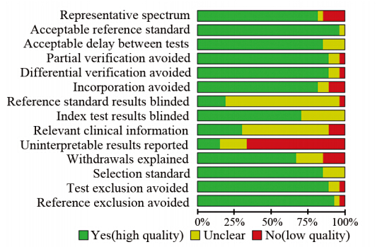 |
| 图 1 纳入PET/CT诊断宫颈癌淋巴结转移文献的质量评价Figure 1 Quality assessment of included studies of PET/CT diagnosis of metastatic lymph nodes from cervical cancer |
26个研究(3 5组数据)间存在异质性(P=0.0000,I2=78.4%),采用随机效应模型进行Meta分析,结果显示:SEN合并、SPE合并、+LR合并、-LR合并、DOR、AUROC分别为0.73(95% CI:0 .7 0 ~0 .7 6),0 .9 6(9 5 % CI:0 .9 5 ~0 .9 6),7 .8 6 ( 9 5 % CI:5 .3 0 ~11 .6 5 ),0 .3 5(9 5 % CI:0.28~0.45),31.63(95% CI:17.17~58.25),0.92(95% CI: 0.89~0.94),见图 2,验前概率和验后概率分别为20%和73%,见图 3。由于卡方检验结果显示各研究间存在高度异质性,使用二元箱线图识别存在异质性的研究,二元箱线图内圈的椭圆呈现了处于中位数分布区域的研究,外圈的椭圆是95%可信区间(CI);可以提供阈值异常的间接证据。PET/CT对宫颈癌淋巴结转移的诊断性能具有中度的敏感度和较好的特异性,大部分的研究都处于中位数分布区域,4组数据分布在95%CI以外,见图 4。排除分布在95%CI以外的4组数据,卡方检验结果显示其余28组数据间异质性减小(P=0.0006,I2=68.8%)。
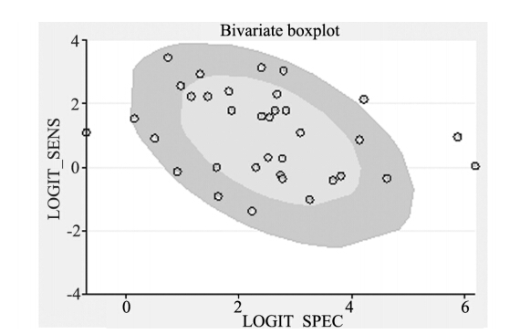 |
| 图 4 纳入35组数据特异性敏感度二元箱线图 Figure 4 Bivariate boxplot of sensitivity and specificity of 35 groups of included data |
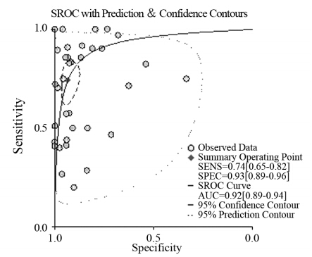 |
| 图 2 纳入35组数据SROC曲线图 Figure 2 SROC curves of included studies |
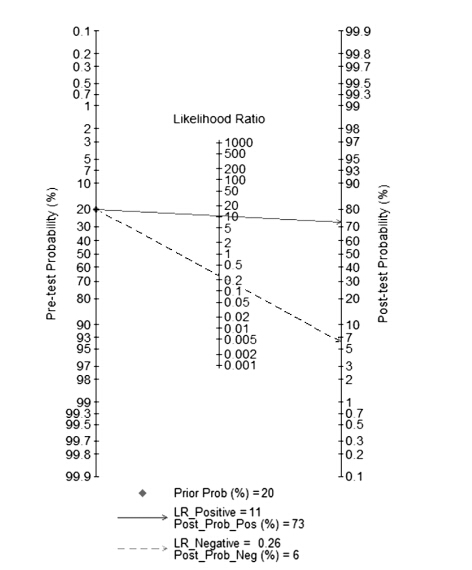 |
| 图 3 PET/CT的验前概率和验后概率计算图 Figure 3 Calculation chart of pre- and post-test probability of PET/CT |
以患者为分析单位,23个研究间有统计学异质性(P=0.0000,I2=70.0%),采用随机效应模型。以淋巴结为分析单位,13个研究间存在统计学异质性(P=0.0000,I2=85.4%),采用随机效应模型,Meta分析结果见表 2。
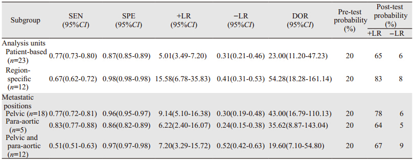 |
根据淋巴结转移部位的不同,18个研究出现盆腔转移,5个研究出现腹主动脉转移,12个研究同时出现了腹主动脉旁和盆腔淋巴结转移。卡方检验结果显示各亚组内研究间存在统计学异质性(P<0.1,I2>50%),采用随机效应模型合并数据,见表 2。2.6 敏感度分析和发表偏倚
以DOR(ln DOR)为横坐标,有效样本量的平方根的倒数(1/ESS1/2)为纵坐标绘制漏斗图,斜率系数P=0.364[t=-0.92,95% CI(-17.35,6.55)],说明存在发表偏倚可能性较小,见图 5。
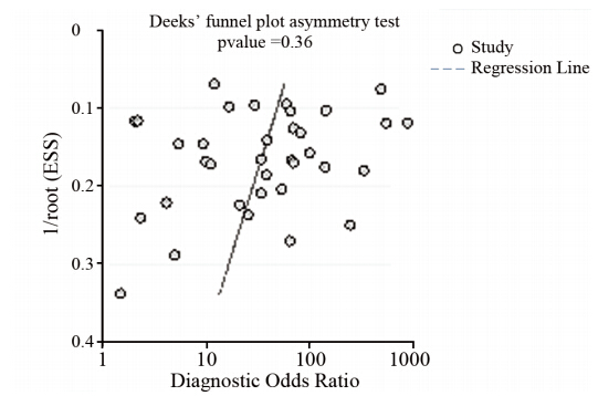 |
| 图 5 纳入35组数据PET/CT的Deek’s 漏斗图 Figure 5 Deek’s funnel plot of PET/CT of 35 groups of included dataincluded data |
宫颈癌是全球妇女中仅次于乳腺癌和结直肠癌的第3个常见的恶性肿瘤,也是最常见的女性生殖道恶性肿瘤,随着发病年龄的年轻化,其发生率及治疗后复发率的增加,宫颈癌越来越多的影响着女性的健康,因此对其尽早准确诊断及分析淋巴结转移具有重要的临床意义。本系统评价纳入了26个以手术病理检查作为参考标准的PET/CT诊断宫颈癌淋巴结转移的试验,数据表明其诊断宫颈癌淋巴结转移有中度的敏感度和较好的特异性,可作为宫颈癌淋巴结转移的有效诊断方法之一。
PET/CT在诊断宫颈癌淋巴结转移的敏感度为73%,特异性为96%,但因仍存在27%的漏诊率和4%的误诊率,我们仍不能因为PET/CT显示阴性而排除淋巴结清扫,由于潜在偏倚的存在,PET/CT仍不能够彻底的取代淋巴结清扫术。但是对于已经失去做淋巴结清扫机会的患者,PET/CT诊断可帮助临医生选择合适的治疗方式,以期提高患者生存质量和5年生存率[36]。PET/CT诊断宫颈癌淋巴结转移的特异性较高,敏感度中等,可能与PET/CT对于直径<0.5 cm的淋巴结转移存在一定的假阴性有关。亚组分析结果显示,以患者为分析单位较以淋巴结为分析单位具有更高的敏感度,且阳性似然比的验后概率明显高于验前概率,提示当PET/CT诊断为阳性时,可有效地对宫颈癌淋巴结转移疑似病例的筛查。以淋巴结为分析单位较以患者为分析单位具有更高的特异性、似然比和诊断比值比,其阴性似然比的验后概率较验前概率提高很多,表明当PET/CT诊断为阴性时,可有效地排除宫颈癌淋巴结转移病例。对淋巴结转移部位进行亚组分析,结果显示了PET/CT对淋巴结盆腔转移的诊断价值优于腹主动脉转移。
本系统评价最终纳入研究间的质量差异不是很大,但高质量研究少,同时由于本文所纳入的文献大多属于回顾性研究,可能存在固有的选择偏倚。本研究存在较大的异质性,合并分析结果显示I2均大于50%,原因可能为PET/CT的判读标准的不一致和患者情况的不同。在文献筛选过程中我们发现:(1)大部分研究在测量结果时没有采用盲法。可能存在解读偏倚;(2)多数文献未报道敏感度和特异性;以上问题势必影响文献的质量和合并分析。
本Meta分析存在一定的局限性:(1)纳入文献的研究对象代表性有限;(2)大多数研究未采用盲法;(3)大多数研究未报告PET/CT检测与手术后病理检测间隔时间;(4)纳入研究样本量较小。因此建议今后的研究:(1)尽量遵循诊断性试验报告标准(STARD),提高诊断性试验的报告质量; (2)诊断性试验和参照标准应尽量同步进行,在进行诊断时应做到“盲法”评估;(3)应详细描述研究对象的特征、试验的方法、质量控制、试验的测量指标等,以便于试验的重现或将试验结果应用于实践;(4)文献中应报告原始数据、计算有关的诊断性试验评价指标及其可信区间。以便为其实际应用和医疗决策提供参考,注意报告因诊断试验或参考试验不可行或结果不确定而被排除的研究对象数;(5)应尽可能采用前瞻性研究方法;(6)对PET/CT用于诊断宫颈癌淋巴结转移,除评价其诊断准确性外,还应当关注安全性和成本一效果等问题。
综上所述,PET/CT在宫颈癌盆腔淋巴结转移方面的诊断能力具有中度的敏感度和较高的特异性,其检查结果有助于我们对早期宫颈癌患者准确地进行术前评估和手术计划的制定,可作为早期宫颈癌患者术前评估的重要辅助手段。
| [1] | Parkin DM, Bray F, Ferlay J, et al. Estimating the world cancer burden:Globocan 2000[J]. Int J Cancer, 2001, 94(2): 153-6. |
| [2] | Tanaka Y, Sawada S, Murata T. Relationship between lymph node metastases and prognosis in patients irradiated postoperatively for carcinoma of the uterine cervix[J]. Acta Radiol Oncol, 1984, 23(6): 455-9. |
| [3] | Downey GO, Potish RA, Adcock LL, et al. Pretreatment surgical staging in cervical carcinoma:therapeutic efficacy of pelvic lymph node resection[J]. Am J Obster Gynecol, 1989, 160(5 Pt 1): 1055-61. |
| [4] | Potish RA, Twiggs LB, Okagaki T, et al. Therapeutic implications of the natural history of advanced cervical cancer as defined by pretreatment surgical staging [J].Cancer, 1985, 56(4): 956-60. |
| [5] | Chou HH, Chang HP, Lai CH, et al. (18)F-FDG PET in Stage 1B/ IIB cervical adenocarcinoma/ adenosquamous carcinoma [J]. Eur J Nucl Med Mol Imaging, 2010, 37(4): 728-35. |
| [6] | Grigsby PW. PET/CT imaging to guide cervical cancer therapy[J]. Future Oncol, 2009, 5(7): 953-8. |
| [7] | Dolezelova H, Slampa P, Ondrova B, et al. The impact of PET with 18F-FDG in radio therapy treatment planning and in the prediction inpatient swith cervix carcinoma: results of pilot study[J]. Neoplasma, 2008, 55(5): 437-41. |
| [8] | Leeflang MM, Deeks JJ, Gatsonis C, et al. Systematic reviews of diagnostic test accuracy[J]. Ann Intern Med, 2008, 149(12): 889-97. |
| [9] | Whiting P, Rutjes AW, Reitsma JB, et al. The development of QUADAS: a tool for the quality assessment of studies of diagnostic accuracy included in systematic reviews[J]. BMC Med Res Methodol, 2003, 3: 25. |
| [10] | Rose PG, Adler LP, Rodriguez M, et al. Positron emission tomography for evaluating para-aortic nodal metastasis in locally advanced cervical cancer before surgical staging: a surgicopathologic study[J]. J Clin Oncol, 1999, 17(1): 41-5. |
| [11] | Narayan K, Hicks RJ, Jobling T, et al. A comparison of MRI and PET scanning in surgically staged loco‐regionally advanced cervical cancer: Potential impact on treatment[J]. Int J Gynecol Cancer, 2001, 11(4): 263-71. |
| [12] | Reinhardt MJ, Ehritt-Braun C, Vogelgesang D, et al. Metastatic lymph nodes in patients with cervical cancer: detection with MR imaging and FDG PET[J]. Radiology, 2001, 218(3): 776-82. |
| [13] | Yeh LS, Hung YC, Shen YY, et al. Detecting para-aortic lymph nodal metastasis by positron emission tomography of 18F-fluorodeoxyglucose in advanced cervical cancer with negative magnetic resonance imaging findings[J]. Oncol Rep, 2002, 9(6): 1289-92. |
| [14] | Belhocine T, Thille A, Fridman V, et al. Contribution of wholebody 18FDG PET imaging in the management of cervical cancer[J]. Gynecol Oncol, 2002, 87(1): 90-7. |
| [15] | Ryu SY, Kim MH, Choi SC, et al. Detection of early recurrence with 18F-FDG PET in patients with cervical cancer[J]. J Nucl Med, 2003, 44(3): 347-52. |
| [16] | Havrilesky LJ, Wong TZ, Secord AA, et al. The role of PET scanning in the detection of recurrent cervical cancer[J]. Gynecol Oncol, 2003, 90(1): 186-90. |
| [18] | Lv SM. Clinical value of positron emission tomography and computed tomography (PET/CT) in the monitoring gynecologic malignant tumors[D]. Shandong University, 2005. [吕世明. 正电子发射体层显像 (PET/CT) 检测妇科恶性肿瘤的临床价值[D]. 山东大学, 2005.] |
| [19] | Park W, Park YJ, Huh SJ, et al. The usefulness of MRI and PET imaging for the detection of parametrial involvement and lymph node metastasis in patients with cervical cancer[J]. Jpn J Clin Oncol, 2005, 35(5): 260-4. |
| [20] | Roh JW, Seo SS, Lee S, et al. Role of positron emission tomography in pretreatment lymph node staging of uterine cervical cancer: a prospective surgicopathologic correlation study[J]. Eur J Cancer, 2005, 41(14): 2086-92. |
| [21] | Choi HJ, Roh JW, Seo SS, et al. Comparison of the accuracy of magnetic resonance imaging and positron emission tomography/ computed tomography in the presurgical detection of lymph node metastases in patients with uterine cervical carcinoma: a prospective study[J]. Cancer, 2006, 106(4): 914-22. |
| [22] | Sironi S, Buda A, Picchio M, et al. Lymph node metastasis in patients with clinical early-stage cervical cancer: detection with integrated FDG PET/CT[J]. Radiology, 2006, 238(1): 272-9. |
| [23] | Chung HH, Jo H, Kang WJ, et al. Clinical impact of integrated PET/CT on the management of suspected cervical cancer recurrence[J]. Gynecol Oncol, 2007, 104(3): 529-34. |
| [24] | Loft A, Berthelsen AK, Roed H, et al. The diagnostic value of PET/CT scanning in patients with cervical cancer: a prospective study[J]. Gynecol Oncol, 2007, 106(1): 29–34. |
| [25] | Yildirim Y, Sehirali S, Avci ME, et al. Integrated PET/CT for the evaluation of para-aortic nodal metastasis in locally advanced cervical cancer patients with negative conventional CT findings[J]. Gynecol Oncol, 2008, 108(1): 154-9. |
| [26] | Lu BY, Che WY, Yao SZ. Role of 18F-FDG PET/CT in diagnosing idiopathy and detecting lymph node metastasis in pelves of cervical cancer[J]. Yi Xue Ying Xiang Xue Za Zhi, 2008, 18(9): 1052-4.[卢宝彦, 车文颖, 姚树展, 等. PET/CT显像在宫颈癌原发灶诊断及盆腔淋巴结分期的价值[J]. 医学影像学杂志, 2008, 18(9): 1052-4.] |
| [27] | Kim SK, Choi HJ, Park SY, et al. Additional value of MR/PET fusion compared with PET/CT in the detection of lymph node metastases in cervical cancer patients[J]. Eur J Cancer, 2009, 45(12): 2103-9. |
| [28] | Kitajima K, Murakami K, Yamasaki E, et al. Accuracy of integrated FDG-PET/contrast-enhanced CT in detecting pelvic and paraaortic lymph node metastasis in patients with uterine cancer[J].Eur Radiol, 2009, 19(6): 1529-36. |
| [29] | Chung HH, Park NH, Kim JW, et al. Role of integrated PET-CT in pelvic lymph node staging of cervical cancer before radical hysterectomy[J]. Gynecol Obstet Invest, 2009, 67(1): 61-6. |
| [30] | Chung HH, Kang KW, Cho JY, et al. Role of magnetic resonance imaging and positron emission tomography/computed tomography in preoperative lymph node detection of uterine cervical cancer[J]. Am J Obstet Gynecol, 2010, 203(2): 156.e1-5. |
| [31] | Sandvik RM, Jensen PT, Hendel HW, et al. Positron emission tomography-computed tomography has a clinical impact for patients with cervical cancer[J]. Dan Med Bull, 2011, 58(3): A4240. |
| [32] | Leblanc E, Gauthier H, Querleu D, et al. Accuracy of 18-fluoro-2-deoxy-D-glucose positron emission tomography in the pretherapeutic detection of occult para-aortic node involvement in patients with a locally advanced cervical carcinoma[J]. Ann Surg Oncol, 2011, 18(8): 2302-9. |
| [33] | Signorelli M, Guerra L, Montanelli L, et al. Preoperative staging of cervical cancer: is 18-FDG-PET/CT really effective in patients with early stage disease?[J]. Gynecol Oncol, 2011, 123(2): 236-40. |
| [34] | Crivellaro C, Signorelli M, Guerra L, et al. 18F-FDG PET/CT can predict nodal metastases but not recurrence in early stage uterine cervical cancer[J]. Gynecol Oncol, 2012, 127(1): 131-5. |
| [35] | Chen HY, Wang DB, Li YN. Diagnostic value of PET-CT in detection of lymph node metastasis in early-stage cervical carcinoma[J]. Zhongguo Yi Ke Da Xue Xue Bao, 2013, 42(4): 351-4.[陈英汉, 王丹波, 李雅男, 等. PET-CT在检测早期子宫颈癌淋巴结转移中的诊断价值[J]. 中国医科大学学报, 2013, 42(4): 351-4.] |
| [36] | Coleman RL, Whitten CW, O'Boyle J, et al. Unexplained decrease in measured oxygen saturation by pulse oximetry following injection of Lymphazurin 1% (isosulfan blue) during a lymphatic mapping procedure[J]. J Surg Oncol, 1999, 70(2): 126-9. |
 2015, Vol. 42


