-
摘要:目的
探讨SEC61G的表达与口腔鳞状细胞癌(OSCC)临床病理特征的相关性及其预后诊断价值。
方法利用TCGA数据库初步分析SEC61G在OSCC中的表达及其预后诊断价值;免疫组织化学检测SEC61G在64对OSCC石蜡组织及其癌旁组织中的表达,并分析SEC61G的表达与OSCC临床病理特征的相关性;Kaplan-Meier法分析SEC61G的表达与OSCC患者术后总体生存期(OS)的关系;Cox回归分析影响OSCC患者预后的因素。
结果SEC61G在OSCC组织中的表达明显高于癌旁组织(P < 0.05),与TCGA数据库结果一致,且其表达与OSCC患者的N分期、临床分期相关(P < 0.05);SEC61G低表达组中OSCC患者的总体生存期明显高于SEC61G高表达组(P < 0.05);N分期、SEC61G的表达均是OSCC患者预后的影响因素,且SEC61G的表达是独立预测因素(P < 0.05);ROC曲线下面积为0.923。
结论SEC61G在OSCC组织中高表达,与患者N分期、临床分期相关,且其高表达与患者预后不良有关,可能是OSCC的一个诊断标志物。
Abstract:ObjectiveTo analyze the expression of SEC61G in oral squamous cell carcinoma (OSCC) tissues and cell lines and determine its correlations with the clinicopathological features and prognosis of patients with OSCC.
MethodsThe expression of SEC61G in OSCC tissues and its diagnostic and prognostic value were detected in the TCGA database. The expression levels of SEC61G in paraffin-embedded OSCC tissues and adjacent normal tissue specimens of 64 patients with OSCC were detected by immunohistochemistry. The correlation of SEC61G expression in OSCC tissues with the clinicopathological features was analyzed. Kaplan-Meier survival curve analysis showed that the expression of SEC61G correlated with the overall survival time of patients with OSCC. Cox regression analysis was used to analyze prognostic influence factors.
ResultsThe expression of SEC61G in OSCC tissues was significantly higher than that in para-carcinoma tissues (P < 0.05), consistent with the results of the TCGA database analysis, and its expression was closely related to N stage and clinical stage (P < 0.05). The overall survival of patients with OSCC in the group with low SEC61G expression was significantly higher than that in the high SEC61G expression group (P < 0.05). N stage and SEC61G expression were the prognostic influence factors for patients with OSCC (P < 0.05). SEC61G expression was an independent predictor of the prognosis of patients with OSCC (P < 0.05, AUC=0.923).
ConclusionSEC61G is highly expressed in OSCC tissues and associated with N stage and clinical stage. Its high expression is associated with poor prognosis of patients. It may be a diagnostic biomarker for OSCC.
-
Key words:
- Oral squamous cell carcinoma /
- SEC61G /
- Clinicopathological features /
- Prognosis /
- Diagnosis
-
0 引言
口腔鳞状细胞癌(oral squamous cell carcinoma, OSCC)起源于口腔上皮组织,是最常见的颌面部恶性肿瘤。据统计,OSCC占口腔恶性肿瘤的90%以上,全球大约每年新增500 000例OSCC患者[1],以40~60岁的男性患者多见,且其发病率随年龄的增长而上升[2]。烟、酒、槟榔等均是OSCC的致病因素,且近些年发现口腔微生物群与OSCC的发生发展密切相关[3]。OSCC具有恶性程度高、局部浸润性强、淋巴结转移率高等特点,晚期可发生远处转移,其5年生存率约55%[4-5]。尽管临床上针对OSCC的手术及放化疗等治疗方法不断改进,但其预后并没有明显的好转,仍缺乏有效的生物标志物[6]。因此,发现OSCC特异性标志物可能会提高OSCC患者的生存率,改善其预后状况。
SEC61G(SEC61 translocon subunit gamma, SEC61G)本质上是一种细胞膜蛋白,该膜蛋白是SEC61复合体的重要成员之一[7]。SEC61复合体是由SEC61α、β、γ三个亚基形成的异质三聚体蛋白通道,是细胞内质网膜蛋白转运装置的核心组件[8]。多项研究表明,SEC61G在胶质母细胞瘤[9]、胃癌[10]、肝癌[11]等恶性肿瘤中表达失调,并可作为胶质母细胞瘤的潜在预后标志物[9]。然而,SEC61G与OSCC之间的关系尚不清楚。
本研究首先利用TCGA数据库分析了SEC61G在OSCC组织和癌旁组织中的表达情况,用免疫组织化学的方法对结果进行了验证,并进一步分析SEC61G的表达与OSCC患者临床病理特性的相关性,最后探索SEC61G对OSCC的预后和诊断价值,以期为SEC61G作为OSCC诊疗的生物标志物奠定理论基础。
1 资料与方法
1.1 研究对象
本研究于2015—2018年在郑州大学第一附属医院口腔医院收集了64对OSCC石蜡组织及其癌旁组织。纳入标准:(1)有详细的临床病理资料,包括患者的年龄、性别、吸烟史、肿瘤位置、T分期、N分期、临床分期、淋巴结转移、组织分化;(2)术前未行放化疗,均行外科手术治疗且术后配合适当的放化疗;(3)无糖尿病等严重并发症。排除标准:(1)临床病理资料不全;(2)复发病例或术前接受过放化疗;(3)非原发于口腔颌面部的肿瘤。
1.2 TCGA数据库样本
通过TCGA数据库(https://portal.gdc.cancer.gov/)下载了OSCC患者的mRNA表达谱信息,舍弃掉没有临床信息对应的数据,共得到329例OSCC组织和32例正常组织、32对配对的OSCC组织和癌旁组织(格式:TPM,平台:RNAseq)。筛选出SEC61G的表达量和临床资料,利用仙桃学术(https://www.xiantaozi.com/)在线分析软件分析SEC61G在OSCC组织中的表达与患者N分期、临床分期、放疗史的相关性,并探讨了SEC61G的表达对OSCC的预后、诊断价值,用ggplot2[3.3.6]包对数据进行可视化。
1.3 免疫组织化学染色
采用常规免疫组织化学法检测SEC61G在OSCC组织及配对癌旁组织中的表达。具体步骤如下:在切片机上制备5 μm蜡块切片,60℃烤片2 h,24 h后4℃保存;经脱蜡、水化后,在每张切片上加50 μl的过氧化物酶阻断剂,室温条件下孵育10 min,PBS洗3次,每次5 min;置于1 000 ml EDTA(pH=8.0的乙二胺四乙酸)抗原修复液中修复抗原,PBS缓冲液浸泡、洗涤3次,约5分钟/次,PBST冲洗5 min;50 μl封闭液封闭,滴加一抗、二抗;显色,苏木精对比染色,梯度酒精(50%、75%、95%、100%)脱水,中性树脂封片,拍照。
染色结果的判读:由两位病理科医生采用双盲法评估,分别记录切片中SEC61G阳性细胞的比例和染色强度。染色强度分级如下:0: 未着色;1: 浅黄色;2: 棕黄色;3: 棕褐色。染色面积分级如下:0: ≤5%;1: > 5%~25%;2: > 25%~50%;3: > 50%~75%;4: > 75%~100%。 < 3分为SEC61G低表达,≥3分为SEC61G高表达。根据两者的得分之和判断SEC61G的表达高低。如果两名医师的判定结果不一致,则由第三名医师进行阅片并确定最终结果。
1.4 随访
对纳入的所有OSCC患者通过病历查阅、门诊复诊、电话致电的方式进行随访并记录患者的生存状态和总生存期,确保所有患者均接受3年以上的随访时间,中途失访者的生存期以最后一次随访时间为准。本研究从2016年6月开始随访,截止时间为2021年9月,每半年随访一次。
1.5 统计学方法
使用SPSS23.0版进行统计分析,所有数值均使用均数±标准差表示。卡方检验用于分析SEC61G的表达与OSCC患者临床病理特性的相关性。为了分析SEC61G的表达水平与OSCC患者预后的关系,根据免疫组织化学的结果,将64对OSCC石蜡标本分为SEC61G低表达和高表达组,并结合患者的临床随访资料,使用Kaplan-Meier法分析OSCC患者3年随访的生存资料,Log rank法比较SEC61G高表达和低表达组中OSCC患者总体生存期的差异。单因素和多因素Cox回归分析影响OSCC患者预后的危险因素。P < 0.05表示差异具有统计学意义。
2 结果
2.1 TCGA数据库中SEC61G在OSCC中的表达
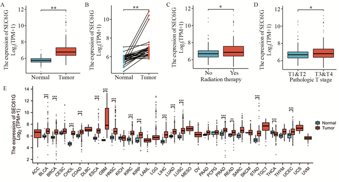
在TCGA数据库中,SEC61G在OSCC组织中的表达明显高于正常组织(P=0.005),见图 1A;在32对配对的OSCC组织和癌旁组织中,SEC61G在肿瘤组织中的表达也明显增加(P=0.003),见图 1B;SEC61G在OSCC中的表达与患者T分期(P= 0.047)、放疗史(P=0.036)相关,见图 1C~D。此外,SEC61G在乳腺癌、前列腺癌等大多数恶性肿瘤的表达均显著增加(P < 0.05),见图 1E。
![]() 图 1 TCGA数据库中SEC61G在OSCC和其他恶性肿瘤组织中的表达Figure 1 Expression of SEC61G in OSCC tissues and other tumor tissues obtained from the TCGA databaseA: The expression of SEC61G mRNA in OSCC cancer tissues (n=329) and normal tissues (n=32); B: The expression of SEC61G mRNA in 32 pairs of OSCC tissues and adjacent normal tissues; C: The relationship between the expression of SEC61G and radiation therapy; D: The relationship between the expression of SEC61G and T classification; E: The expression of SEC61G mRNA in other tumor tissues. *: P < 0.05, **: P < 0.01, ***: P < 0.001.
图 1 TCGA数据库中SEC61G在OSCC和其他恶性肿瘤组织中的表达Figure 1 Expression of SEC61G in OSCC tissues and other tumor tissues obtained from the TCGA databaseA: The expression of SEC61G mRNA in OSCC cancer tissues (n=329) and normal tissues (n=32); B: The expression of SEC61G mRNA in 32 pairs of OSCC tissues and adjacent normal tissues; C: The relationship between the expression of SEC61G and radiation therapy; D: The relationship between the expression of SEC61G and T classification; E: The expression of SEC61G mRNA in other tumor tissues. *: P < 0.05, **: P < 0.01, ***: P < 0.001.2.2 免疫组织化学检测SEC61G在OSCC组织和癌旁组织中的表达
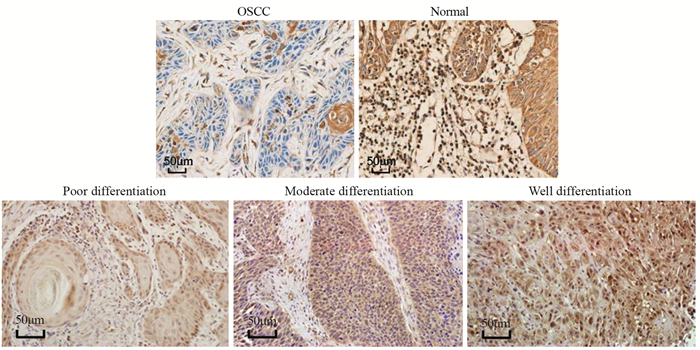
与癌旁组织相比,OSCC组织中SEC61G蛋白的阳性表达率增加,差异具有统计学意义(P=0.031)。SEC61G在低分化OSCC组织中表达最高,在高分化OSCC组织中表达最低,见图 2。
2.3 SEC61G的表达与OSCC患者临床病理特征的相关性
根据免疫组织化学的结果,分为SEC61G低表达和高表达组,并与患者的临床病理资料进行相关性分析。结果表明,SEC61G的表达与OSCC患者N分期、临床分期相关(P < 0.05),见表 1。
表 1 SEC61G的表达与OSCC患者临床病理特征的相关性Table 1 Relationship between SEC61G expression and clinicopathological features of patients with OSCC
2.4 SEC61G表达与OSCC患者预后不良的相关性
SEC61G低表达组中OSCC患者的总体生存率明显高于SEC61G高表达组(P=0.011),见图 3A。此外,在TCGA数据库中,SEC61G低表达组中OSCC患者的总体生存期(P=0.008)、疾病特异性生存期(P=0.002)和无进展生存期(P < 0.001)均显著高于SEC61G高表达组,见图 3B~D。上述结果提示,SEC61G高表达与OSCC患者预后不良有关。
![]() 图 3 SEC61G高表达与OSCC患者预后不良有关Figure 3 High SEC61G expression was associated with poor prognosis of patients with OSCCA: Kaplan-Meier survival curve was used to analyze the relationship between the expression of SEC61G in 64 paraffin specimens and the overall survival of patients with OSCC. The relationship between the expression of SEC61G in the TCGA database and overall survival (B), disease-specific survival (C), and progression-free survival (D) of patients with OSCC.
图 3 SEC61G高表达与OSCC患者预后不良有关Figure 3 High SEC61G expression was associated with poor prognosis of patients with OSCCA: Kaplan-Meier survival curve was used to analyze the relationship between the expression of SEC61G in 64 paraffin specimens and the overall survival of patients with OSCC. The relationship between the expression of SEC61G in the TCGA database and overall survival (B), disease-specific survival (C), and progression-free survival (D) of patients with OSCC.2.5 Cox回归分析影响OSCC患者预后的因素
Cox单因素分析结果表明,N分期和SEC61G的表达是OSCC患者总体生存期的影响因素;多因素分析结果表明,SEC61G的表达是影响OSCC患者预后的独立因素(P < 0.05),见表 2。
表 2 单因素和多因素Cox回归分析影响OSCC患者预后的因素Table 2 Univariate and multivariate Cox analysis of prognostic factors for patients with OSCC
2.6 SEC61G对OSCC患者的诊断价值
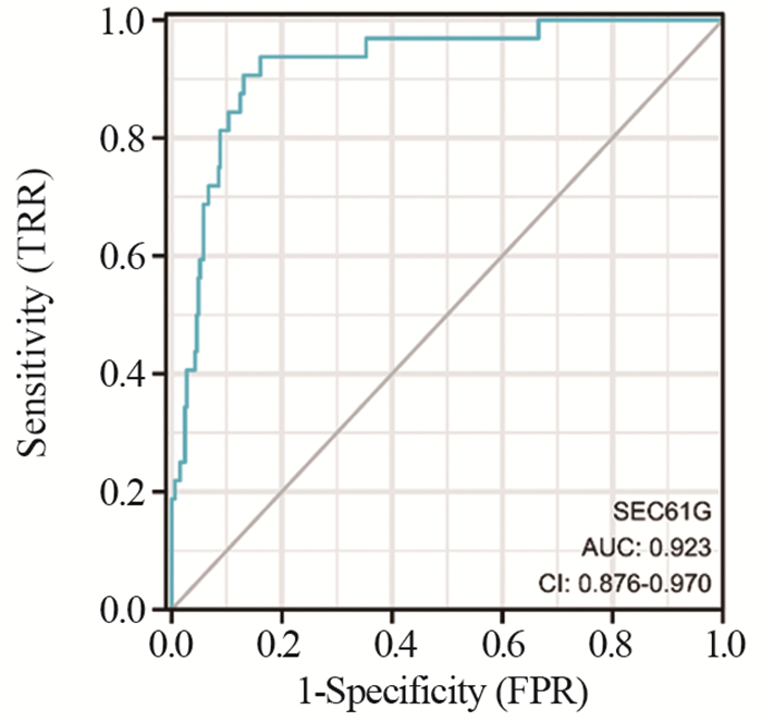
在TCGA数据库中分析了SEC61G对OSCC患者的诊断意义,结果显示,ROC曲线下面积大于0.9(AUC=0.923),表明SEC61G的表达对于OSCC患者的诊断有较高的准确性,可能是OSCC的一个诊断标志物。
3 讨论
OSCC是由生物、化学、遗传等因素引起的口腔颌面部常见的恶性肿瘤,近些年发病率逐渐增高,一旦发生转移预后往往较差,严重影响患者的生命健康[12]。由于OSCC早期症状不明显,发现时多为中晚期,增加了治疗难度,病死率较高,因此早期诊断对于提高OSCC患者的生存期尤为重要。从基因水平检测与恶性肿瘤相关的蛋白或RNA,发现潜在的生物标志物,已经被证明对于提高肿瘤患者的诊疗方法、改善患者的预后状况均有重要意义[13]。
SEC61是一种内质网膜转运复合体,定位于内质网膜,对新合成的前体多肽运输到内质网至关重要[8]。除了SEC61外,由SEC62和SEC63形成的完整的蛋白质转位酶也参与蛋白质的折叠、翻译后修饰和转运[7, 14]。研究表明,内质网通道蛋白与肿瘤的发生发展有关,例如SEC62和SEC63在多种恶性肿瘤中发生突变或表达异常[15-16]。然而,关于SEC61和恶性肿瘤的研究较少。SEC61G是SEC61的三个亚基之一,据报道,SEC61G在多形性胶质母细胞瘤中的表达显著增加,促进肿瘤细胞增殖,并参与内质网应激[9]。Gao等研究表明SEC61G在肝癌组织中的表达显著高于癌旁组织,且其表达水平与肝癌患者的总体生存期相关,可能是肝癌的一个治疗靶点[11]。另有研究表明,SEC61G在肾癌组织中表达失调,敲低SEC61G可促进肾癌细胞凋亡和上皮-间充质转化(EMT)[17]。Zheng等通过生物信息学分析发现SEC61G在肺腺癌中表达上调,与肺腺癌患者预后不良有关,且降低SEC61G表达可抑制乳腺癌细胞的增殖、迁移、侵袭、促进其凋亡[18],Xu等的研究也得到同样的结果[19]。Jin等研究表明降低SEC61G表达可抑制乳腺癌细胞的增殖、迁移、侵袭、促进其凋亡,并可诱导EMT[20]。此外,Ma等研究发现SEC61G通过调节糖酵解促进乳腺癌的发生发展,可能是乳腺癌的一个潜在治疗靶点[21]。然而,SEC61G在OSCC中的作用并不清楚。
针对恶性肿瘤中差异基因的表达和预后分析,有助于发现新的肿瘤标志物。在本研究中,首先初步通过TCGA数据库分析了SEC61G在OSCC组织和正常组织中的表达,结果发现SEC61G在OSCC组织中的表达明显上调,与患者N分期、临床分期、放疗史相关,并且在前列腺癌和胆管癌等大多数恶性肿瘤中的表达均显著增高,表明SEC61G可能是一个癌基因。然后,进一步通过免疫组织化学检测了SEC61G在64例OSCC石蜡组织及其配对癌旁组织中的表达,根据其表达水平分为SEC61G高表达和低表达组,并与患者临床病理特征进行相关性分析,结果显示SEC61G在OSCC组织中的表达显著高于癌旁组织,且其表达与患者N分期、临床分期相关,再次验证了TCGA数据库的分析结果。同时,我们发现SEC61G低表达组中OSCC患者的总体生存期、疾病特异性生存期和无进展生存期均显著高于SEC61G高表达组,提示SEC61G高表达与OSCC患者预后不良有关。此外,Cox回归分析的结果显示SEC61G的表达是OSCC患者预后的独立影响因素。最后,利用TCGA数据库中的临床资料分析SEC61G对OSCC的诊断价值,结果表明SEC61G有作为OSCC诊断标志物的潜力。
综上,SEC61G在OSCC组织中高表达,且其高表达与OSCC患者预后不良有关,可能是OSCC的一个预后、诊断标志物。然而,本研究也存在一定的不足之处,比如临床样本量较少且缺乏相关的细胞功能学实验。未来可联合多中心增加临床样本量,并进一步探讨SEC61G影响OSCC发生、发展的分子机制。
Competing interests: The authors declare that they have no competing interests.利益冲突声明:所有作者均声明不存在利益冲突。作者贡献:高瑞:研究设计、资料收集、论文撰写郭磊:资料收集彭博:统计分析贾骏:研究设计、论文撰写指导 -
表 1 SEC61G的表达与OSCC患者临床病理特征的相关性
Table 1 Relationship between SEC61G expression and clinicopathological features of patients with OSCC

表 2 单因素和多因素Cox回归分析影响OSCC患者预后的因素
Table 2 Univariate and multivariate Cox analysis of prognostic factors for patients with OSCC

-
[1] Miller KD, Nogueira L, Mariotto AB, et al. Cancer treatment and survivorship statistics, 2019[J]. CA Cancer J Clin, 2019, 69(5): 363-385. doi: 10.3322/caac.21565
[2] Siegel RL, Miller KD, Fuchs HE, et al. Cancer Statistics, 2021[J]. CA Cancer J Clin, 2021, 71(1): 7-33. doi: 10.3322/caac.21654
[3] Stasiewicz M, Karpiński TM. The oral microbiota and its role in carcinogenesis[J]. Semin Cancer Biol, 2022, 86(Pt 3): 633-642.
[4] Ferlay J, Soerjomataram I, Dikshit R, et al. Cancer incidence and mortality worldwide: sources, methods and major patterns in GLOBOCAN 2012[J]. Int J Cancer, 2015, 136(5): E359-E386.
[5] Torre LA, Siegel RL, Ward EM, et al. Global Cancer Incidence and Mortality Rates and Trends--An Update[J]. Cancer Epidemiol Biomarkers Prev, 2016, 25(1): 16-27. doi: 10.1158/1055-9965.EPI-15-0578
[6] Mumtaz M, Bijnsdorp IV, Böttger F, et al. Secreted protein markers in oral squamous cell carcinoma (OSCC)[J]. Clin Proteomics, 2022, 19(1): 4. doi: 10.1186/s12014-022-09341-5
[7] Linxweiler M, Schick B, Zimmermann R. Let's talk about Secs: Sec61, Sec62 and Sec63 in signal transduction, oncology and personalized medicine[J]. Signal Transduct Target Ther, 2017, 2: 17002. doi: 10.1038/sigtrans.2017.2
[8] Greenfield JJ, High S. The Sec61 complex is located in both the ER and the ER-Golgi intermediate compartment[J]. J Cell Sci, 1999, 112(Pt 10): 1477-1486.
[9] Liu B, Liu J, Liao Y, et al. Identification of SEC61G as a Novel Prognostic Marker for Predicting Survival and Response to Therapies in Patients with Glioblastoma[J]. Med Sci Monit, 2019, 25: 3624-3635. doi: 10.12659/MSM.916648
[10] Tsukamoto Y, Uchida T, Karnan S, et al. Genome-wide analysis of DNA copy number alterations and gene expression in gastric cancer[J]. J Pathol, 2008, 216(4): 471-482. doi: 10.1002/path.2424
[11] Gao H, Niu W, He Z, et al. SEC61G plays an oncogenic role in hepatocellular carcinoma cells[J]. Cell Cycle, 2020, 19(23): 3348-3361. doi: 10.1080/15384101.2020.1843816
[12] Bugshan A, Farooq I. Oral squamous cell carcinoma: metastasis, potentially associated malignant disorders, etiology and recent advancements in diagnosis[J]. F1000Res, 2020, 9: 229. doi: 10.12688/f1000research.22941.1
[13] Cesur-Ergün B, Demir-Dora D. Gene therapy in cancer[J]. J Gene Med, 2023: e3550.
[14] Liu Y, Ji W, Shergalis A, et al. Activation of the Unfolded Protein Response via Inhibition of Protein Disulfide Isomerase Decreases the Capacity for DNA Repair to Sensitize Glioblastoma to Radiotherapy[J]. Cancer Res, 2019, 79(11): 2923-2932. doi: 10.1158/0008-5472.CAN-18-2540
[15] Wemmert S, Lindner Y, Linxweiler J, et al. Initial evidence for Sec62 as a prognostic marker in advanced head and neck squamous cell carcinoma[J]. Onco Lett, 2016, 11(3): 1661-1670. doi: 10.3892/ol.2016.4135
[16] Casper M, Weber SN, Kloor M, et al. Hepatocellular carcinoma as extracolonic manifestation of Lynch syndrome indicates SEC63 as potential target gene in hepatocarcinogenesis[J]. Scand J Gastroenterol, 2013, 48(3): 344-351. doi: 10.3109/00365521.2012.752030
[17] Meng H, Jiang X, Wang J, et al. SEC61G is upregulated and required for tumor progression in human kidney cancer[J]. Mol Med Rep, 2021, 23(6): 427. doi: 10.3892/mmr.2021.12066
[18] Zheng Q, Wang Z, Zhang M, et al. Prognostic value of SEC61G in lung adenocarcinoma: a comprehensive study based on bioinformatics and in vitro validation[J]. BMC Cancer, 2021, 21(1): 1216. doi: 10.1186/s12885-021-08957-4
[19] Xu S, Li X, Geng J, et al. Sec61γ is a vital protein in the endoplasmic reticulum membrane promoting tumor metastasis and invasion in lung adenocarcinoma[J]. Br J Cancer, 2023, 128(8): 1478-1490. doi: 10.1038/s41416-023-02150-z
[20] Jin L, Chen D, Hirachan S, et al. SEC61G regulates breast cancer cell proliferation and metastasis by affecting the Epithelial-Mesenchymal Transition[J]. J Cancer, 2022, 13(3): 831-846. doi: 10.7150/jca.65879
[21] Ma J, He Z, Zhang H, et al. SEC61G promotes breast cancer development and metastasis via modulating glycolysis and is transcriptionally regulated by E2F1[J]. Cell Death Dis, 2021, 12(6): 550. doi: 10.1038/s41419-021-03797-3


 下载:
下载:


