-
摘要:
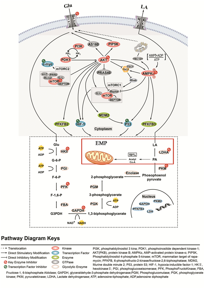
近年来,我国结直肠癌发病率及死亡率逐年上升,寻找早期预防、诊断和治疗的有效途径是目前结直肠癌基础及临床的研究热点。机体正常细胞主要由葡萄糖氧化供能,而结直肠癌细胞的糖酵解途径显著增强并以此作为主要供能途径。本文就糖酵解关键酶、信号转导通路和转录因子在结直肠癌糖酵解途径中的研究进展及治疗策略作一综述。
Abstract:Recently, the incidence and mortality of colorectal cancer in China have been increasing year by year. So nowadays, looking for an effective way to prevent, diagnose and treat early colorectal cancer has been the focus in the clinical and basic research of colorectal cancer. The body's normal cells are mainly oxidized by glucose, while the colorectal cancer cells have an obvious enhanced Embden-Meyerhof-Parnas that serves as a major source of energy. In this paper, the research progress and therapeutic strategies of glycolysis key enzyme, signal transduction pathway and transcription factor in the Embden-Meyerhof-Parnas of colorectal cancer are reviewed.
-
0 引言
目前,食管癌的发病率和死亡率均较高(占恶性肿瘤总发病率的4.2%,总死亡率的6.6%)[1],且我国食管癌的发病率和死亡率均高于全球平均水平[2],放疗是食管癌重要的治疗手段之一,而放射性肺炎(radiation pneumonitis, RP)作为胸部放疗最常见的不良反应,短期内可引起咳嗽、气短、发热等症状,长期可引起肺纤维化、肺功能损伤,影响患者的生活质量,严重时甚至导致患者死亡,且放射性肺炎会限制临床医生给予的处方剂量,从而影响治疗效果和患者预后。RP的发病率也较高,有学者统计发现食管癌患者接受放疗后发生≥2级RP的风险约为22%[3],而老年食管患者接受放疗后发生RP的风险可高达52.4%[4],这提示老年食管癌患者在接受放射治疗时需要更多重视。为避免RP发生,剂量体积直方图(dose-volume histogram, DVH)常用于评估放疗计划,其中肺平均剂量(mean lung dose, MLD)和V20(接受≥20Gy肺体积占总肺体积百分比)常作为约束指标,但效果欠佳[5],且目前应用DVH参数预测放疗后发生RP的研究尚无统一定论[6-8]。因此,本研究分析来自不同中心的老年食管癌患者的剂量体积参数与三维适形放疗后发生≥2级放射性肺炎的相关性,旨在为预防老年患者发生放射性肺炎提供帮助。
1 资料与方法
1.1 临床资料
本研究为回顾性分析,选择2018年1月至2020年1月在东南大学附属中大医院以及江苏省泰州市靖江市人民医院接受三维适形放疗的食管癌患者,收集资料包括:(1)临床特征:性别、年龄、一般体力状况ECOG评分、吸烟史、化疗史。(2)剂量体积参数:V5、V10、V20、V30、MLD。患者放疗开始及放疗期间每两周进行随访,放疗结束后每月进行随访,随访内容包括采集病史、体格检查及胸部CT平扫检查,随访时间为3月。纳入标准:(1)病理明确诊断为食管鳞癌;(2)年龄:60~80岁;(4)PS评分:ECOG 0~2分;(5)放疗期间及结束后完整接受随访(3月),包括病史采集、查体及胸部CT平扫。排除标准:合并肺部基础疾病:包括肺间质性疾病、慢性阻塞性肺疾病等。共收集符合标准的患者250例,其中男151例(60.4%)、女99例(39.6%),平均年龄71岁;东南大学附属中大医院病例110例(44%),靖江市人民医院140例(56%)。
1.2 治疗计划与实施
所有患者均接受三维适形放疗,东南大学附属中大医院应用热塑模固定,定位CT以5 mm层厚扫描,包括中下颈部和全胸部及上腹部,传输图像至Release 4.3.1治疗系统,放疗使用西门子Primus-m直线加速器。靖江市人民医院应用真空垫体模固定,定位CT以3 mm层厚扫描,包括中下颈部和全胸部及上腹部,传输图像至Eclipse 8.6治疗计划系统,放疗使用Varian23 EX直线加速器。食管癌PTV为CTV外扩5~10 mm,根治性放疗处方剂量为60 Gy,术后辅助放疗处方剂量50~60 Gy,均应用2 Gy/F常规分割,以98%等剂量线包绕95%以上计划靶体积,正常组织限量:脊髓剂量 < 45 Gy;心脏V30 < 40%,V40 < 30%;双肺平均剂量 < 20 Gy,V20 < 30%,V30 < 20%,同步化疗时双肺V20限制 < 28%。250例老年食管癌患者中,113例食管鳞癌患者接受了小剂量顺铂/奈达铂同步放化疗(45.2%)。
1.3 放射性肺炎的诊断与分级
放射性肺炎诊断标准采用肿瘤放射治疗学第5版标准:(1)既往6月内有肺受照射病史;(2)CT影像学改变主要局限在照射区域内,病变与正常肺组织的解剖结构不符;(3)多有咳嗽、气短、发热等临床症状;(4)排除能引起类似症状的其他因素[9]。放射性肺炎的分级标准采用不良事件通用术语标准第5版分为:1级:无症状,仅临床或影像学所见,无需治疗;2级:有症状,影响应用工具的日常活动,需治疗;3级:重度症状,影响自理性活动,需吸氧;4级:危及生命的呼吸障碍,需气管切开或插管;5级:死亡[10],由至少两名放疗专科医生评估分级。
1.4 统计学方法
采用SPSS23.0软件对数据进行统计分析,卡方检验分析放射性肺炎组与非放射性肺炎组患者临床特征的差异;单因素Logistic回归分析DVH参数中与发生≥2级RP相关的因素;将单因素分析中有统计学意义的DVH参数纳入多因素Logistic回归,分析与发生≥2级RP独立相关的因素;应用ROC曲线分析发生≥2级RP独立相关的DVH参数的AUC值及最佳分界值,取约登指数最大时的值。样本量采用EPV法(events per variable)确定。P < 0.05为差异有统计学意义。
2 结果
2.1 患者的临床特征及放射性肺炎发生率
将放射性肺炎组与非放射肺炎组患者的临床特征进行卡方检验,两组间性别(P=0.561)、平均年龄(P=0.948)、ECOG评分(P=0.515)、吸烟史(P=0.604)、化疗史(P=0.849)差异均无统计学意义,见表 1。
表 1 250例老年食管癌患者的临床特征及卡方检验结果Table 1 Clinical features and chi-square test results of 250 elderly patients with esophageal cancer
2.2 放射性肺炎相关DVH参数的单因素回归分析结果
Logistic单因素分析与发生≥2级放射性肺炎相关的剂量体积参数,结果提示双肺V5(P < 0.05)、V10(P < 0.05)、V20(P < 0.05)、V30(P < 0.05)及MLD(P < 0.05)均是老年食管癌患者三维适形放疗后发生≥2级RP的相关因素,见表 2。
表 2 250例老年食管癌患者发生≥2级RP的Logistic单因素分析Table 2 Logistic univariate analysis for ≥grade 2 RP in 250 elderly patients with esophageal cancer
2.3 放射性肺炎相关DVH参数的多因素回归分析结果
将单因素分析结果中与RP有显著相关性的DVH参数:双肺V5、V10、V20、V30、MLD进行Logistic多因素分析,结果显示仅双肺V5(P=0.016)、V20(P=0.005)有统计学意义,提示双肺V5、V20是老年食管癌患者三维适形放疗后发生≥2级RP的独立相关因素;双肺V10(P=0.900)、V30(P=0.114)及MLD(P=0.441)与发生≥2级RP有相关性,但不是发生≥2级RP的独立相关因素,见表 3。
表 3 250例老年食管癌患者发生≥2级RP的Logistic多因素分析Table 3 Logistic multivariate analysis for ≥grade 2 RP in 250 elderly patients with esophageal cancer
2.4 ROC曲线分析结果
应用ROC曲线分析V5及V20预测≥2级放射性肺炎的效果及最佳分界值,见图 1。V5的ROC曲线下面积为0.851,95%CI: 0.801~0.902;V20的ROC曲线下面积为0.899,95%CI: 0.850~0.949,见表 4;V5预测≥2级RP的最佳分界值为53.90%,敏感度0.92,特异性0.66,约登指数为0.58;V20的最佳分界值为23.15%,敏感度为0.74,特异性为0.91,约登指数为0.66,见表 5。
表 4 双肺V5和V20的ROC曲线下面积Table 4 Area under ROC curve of bilateral pulmonary V5 and V20 表 5 双肺V5和V20的最佳分界值Table 5 The best cut-off value of bilateral pulmonary V5 and V20
3 讨论
放射性肺炎作为食管癌放疗较常见的并发症之一,是多种细胞和分子相互作用,引起大量成纤维细胞积累、增殖和分化,使细胞外基质沉积过多,最终导致肺纤维化的病理生理过程[11],但是RP发生的具体机制仍未明确,因此传统观点仍将剂量体积参数作为评估放疗计划的主要因素[12],以减少RP的发病风险,但目前尚无统一定论[13];临床工作中则应用V20 < 30%、MLD < 20~23Gy以规避RP的发生,但有研究表明效果欠佳[14];而老年患者的肺功能相对减弱,承受损伤的能力较差[15],在接受放射治疗时需要格外重视,因此本研究主要分析老年食管患者三维适形放疗后与≥2级RP相关的剂量体积参数,以便为控制、预防老年患者发生RP提供帮助。
本研究单因素分析表明双肺V5、V10、V20、V30及MLD均是老年食管癌放疗后发生≥2级RP的相关因素,提示DVH参数与RP发生紧密相关,与多数学者观点相符[16-18]。多因素分析结果示双肺V20是≥2级RP的独立相关因素,与多数学者的观点相符[19-21],且目前临床工作评估放疗计划时常约束双肺V20(低于28%~30%)以规避RP,但本研究则发现双肺V20应当 < 23.2%以规避≥2级RP发生,而Tonison的系统回顾也得出了类似的结论,认为应当将V20控制在23%以下[22]。这表明对于老年患者,剂量体积参数的控制应当更加严格,这可能与老年人的基础肺功能较差、肺生理结构改变有关[23]。另本研究还发现MLD与RP具有相关性,但不是RP的独立相关因素,部分关于食管癌患者发生RP的研究也得出了类似结论[24-26],提示对于食管癌患者,MLD预测RP发生的价值还有待进一步探讨。
本研究多因素分析还发现双肺V5也与≥2级RP独立相关,提示低剂量体积参数在预测RP发生方面具有重要价值。沈文斌等随访了222例接受三维适形放疗的食管癌患者,其中22.1%的患者发生了≥2级RP,回归分析发现V5和V20是RP的重要预测因素[27];Zhao等对68例食管癌患者的回顾分析发现,低剂量体积参数在预测RP方面更为重要[28];杜峰等将247例食管癌患者的V5~V40、MLD、GTV及吸烟指数等临床特征进行多因素回归分析,发现双肺V5是≥1级RP及≥3级RP的独立相关因素[29]。这些研究结果均认同了低剂量体积参数在预测RP方面的价值,但也有部分学者提出了不同观点:Zhao等通过对68例患者的回顾分析认为V30是RP的独立相关因素,而非V5和V20[28],但所研究的样本量较少;姚波等针对食管癌及肺癌患者的一项回顾性研究也发现V30与RP独立相关[30],但其研究样本量少(33例),并且在评估危及器官受量时,肺癌患者的双肺体积需扣除GTV后评估,而食管癌患者不存在此问题,因此将肺癌与食管癌患者的肺部DVH参数混合分析可能会对研究结果造成影响。
本研究则应用EPV法确定样本量,共收集了250例食管癌患者的数据,样本量较上述研究更多,并针对分析了5项与RP相关的DVH参数;且样本来源于不同等级的医疗中心,更具有代表性;且主要针对肺功能更为脆弱、更易发生RP的老年患者,具有临床意义。
综上所述,双肺V5和V20是老年食管癌患者三维适形放疗后发生≥2级放射性肺炎的独立相关因素;对于老年患者,剂量体积参数的约束应当更加严格,应当控制双肺V5 < 53.9%,V20 < 23.2%以规避≥2级放射性肺炎发生。
-
[1] Siegel RL, Miller KD, Jemal A. Cancer statistics, 2018[J]. CA Cancer J Clin, 2018, 68(1): 7-30. doi: 10.3322/caac.v68.1
[2] Ferlay J, Soerjomataram I, Dikshit R, et al. Cancer incidence and mortality worldwide: Sources, methods and major patterns in globocan 2012[J]. Int J Cancer, 2015, 136(5): E359-86. doi: 10.1002/ijc.29210
[3] Chen W, Zheng R, Baade PD, et al. Cancer statistics in china, 2015[J]. CA Cancer J Clin, 2016, 66(2): 115-32. doi: 10.3322/caac.21338
[4] Martinez-Outschoorn UE, Peiris-Pagés M, Pestell RG, et al. Cancer metabolism: A therapeutic perspective[J]. Nat Rev Clin Oncol, 2017, 14(2): 113. http://d.old.wanfangdata.com.cn/NSTLQK/NSTL_QKJJ02923949/
[5] Smith B, Schafer XL, Ambeskovic A, et al. Addiction to coupling of the warburg effect with glutamine catabolism in cancer cells[J]. Cell Rep, 2016, 17(3): 821-36. doi: 10.1016/j.celrep.2016.09.045
[6] Vuoristo KS, Mars AE, Sanders JPM, et al. Metabolic Engineering of TCA Cycle for Production of Chemicals[J]. Trends Biotechnol, 2016, 34(3): 191-7. doi: 10.1016/j.tibtech.2015.11.002
[7] Hammad N, Rosas-Lemus M, Uribe-Carvajal S, et al. The crabtree and warburg effects: Do metabolite-induced regulations participate in their induction?[J]. Biochim Biophys Acta, 2016, 1857(8): 1139-46. doi: 10.1016/j.bbabio.2016.03.034
[8] Gillies RJ, Gatenby RA. Hypoxia and adaptive landscapes in the evolution of carcinogenesis[J]. Cancer Metastasis Rev, 2007, 26(2): 311-7. doi: 10.1007/s10555-007-9065-z
[9] Li C, Zhang G, Zhao L, et al. Metabolic reprogramming in cancer cells: Glycolysis, glutaminolysis, and bcl-2 proteins as novel therapeutic targets for cancer[J]. World J Surg Oncol, 2016, 14(1): 15.
[10] Sun S, Li H, Chen J, et al. Lactic acid: No longer an inert and end-product of glycolysis[J]. Physiology (Bethesda), 2017, 32(6): 453-63. http://www.ncbi.nlm.nih.gov/pubmed/29021365
[11] Bohme I, Bosserhoff AK. Acidic tumor microenvironment in human melanoma[J]. Pigment Cell Melanoma Res, 2016, 29(5): 508-23. doi: 10.1111/pcmr.12495
[12] Song K, Li M, Xu X, et al. Resistance to chemotherapy is associated with altered glucose metabolism in acute myeloid leukemia[J]. Oncol Lett, 2016, 12(1): 334-42. doi: 10.3892/ol.2016.4600
[13] Roberts DJ, Miyamoto S. Hexokinase ii integrates energy metabolism and cellular protection: Akting on mitochondria and torcing to autophagy[J]. Cell Death Differ, 2015, 22(2): 248-57. doi: 10.1038/cdd.2014.173
[14] Qin Y, Cheng C, Lu H, et al. Mir-4458 suppresses glycolysis and lactate production by directly targeting hexokinase2 in colon cancer cells[J]. Biochem Biophys Res Commun, 2016, 469(1): 37-43. doi: 10.1016/j.bbrc.2015.11.066
[15] Kudryavtseva AV, Fedorova MS, Zhavoronkov A, et al. Effect of lentivirus-mediated shrna inactivation of hk1, hk2, and hk3 genes in colorectal cancer and melanoma cells[J]. BMC Genet, 2016, 17(Suppl 3): 156. doi: 10.1186/s12863-016-0459-1
[16] Katagiri M, Karasawa H, Takagi K, et al. Hexokinase 2 in colorectal cancer: A potent prognostic factor associated with glycolysis, proliferation and migration[J]. Histol Histopathol, 2017, 32(4): 351-60. http://www.ncbi.nlm.nih.gov/pubmed/27363977
[17] Clem BF, O'Neal J, Tapolsky G, et al. Targeting 6-phosphofructo-2-kinase (pfkfb3) as a therapeutic strategy against cancer[J]. Mol Cancer Ther, 2013, 12(8): 1461-70. doi: 10.1158/1535-7163.MCT-13-0097
[18] Lv Y, Zhang B, Zhai C, et al. Pfkfb3-mediated glycolysis is involved in reactive astrocyte proliferation after oxygen-glucose deprivation/reperfusion and is regulated by cdh1[J]. Neurochem Int, 2015, 91: 26-33. doi: 10.1016/j.neuint.2015.10.006
[19] Han J, Meng Q, Xi Q, et al. Interleukin-6 stimulates aerobic glycolysis by regulating pfkfb3 at early stage of colorectal cancer[J]. Int J Oncol, 2016, 48(1): 215-24. doi: 10.3892/ijo.2015.3225
[20] Webb BA, Forouhar F, Szu FE, et al. Structures of human phosphofructokinase-1 and atomic basis of cancer-associated mutations[J]. Nature, 2015, 523(7558): 111-4. doi: 10.1038/nature14405
[21] Yan XL, Zhang XB, Ao R, et al. Effects of shrna-mediated silencing of pkm2 gene on aerobic glycolysis, cell migration, cell invasion, and apoptosis in colorectal cancer cells[J]. J Cell Biochem, 2017, 118(12): 4792-803. doi: 10.1002/jcb.v118.12
[22] Dayton TL, Jacks T, Vander Heiden MG. Pkm2, cancer metabolism, and the road ahead[J]. EMBO Rep, 2016, 17(12): 1721-30. doi: 10.15252/embr.201643300
[23] Taniguchi K, Sakai M, Sugito N, et al. Ptbp1-associated microrna-1 and -133b suppress the warburg effect in colorectal tumors[J]. Oncotarget, 2016, 7(14): 18940-52. http://www.ncbi.nlm.nih.gov/pmc/articles/PMC4951342/
[24] Ginés A, Bystrup S, Ruiz de Porras V, et al. Pkm2 subcellular localization is involved in oxaliplatin resistance acquisition in ht29 human colorectal cancer cell lines[J]. PLoS One, 2015, 10(5): e0123830. doi: 10.1371/journal.pone.0123830
[25] Koukourakis MI, Giatromanolaki A, Sivridis E, et al. Lactate dehydrogenase 5 expression in operable colorectal cancer: Strong association with survival and activated vascular endothelial growth factor pathway-a report of the tumour angiogenesis research group[J]. J Clin Oncol, 2006, 24(26): 4301-8. doi: 10.1200/JCO.2006.05.9501
[26] Kim EY, Choi HJ, Park MJ, et al. Myristica fragrans suppresses tumor growth and metabolism by inhibiting lactate dehydrogenase A[J]. Am J Chin Med, 2016, 44(5): 1063-79. doi: 10.1142/S0192415X16500592
[27] Li X, Zhao H, Zhou X, et al. Inhibition of lactate dehydrogenase a by microrna-34a resensitizes colon cancer cells to 5-fluorouracil[J]. Mol Med Rep, 2015, 11(1): 577-82. doi: 10.3892/mmr.2014.2726
[28] Deng D, Sun P, Yan C, et al. Molecular basis of ligand recognition and transport by glucose transporters[J]. Nature, 2015, 526(7573): 391-6. doi: 10.1038/nature14655
[29] Chen T, Ren Z, Ye LC, et al. Factor inhibiting hif1alpha (fih-1) functions as a tumor suppressor in human colorectal cancer by repressing hif1alpha pathway[J]. Cancer Biol Ther, 2015, 16(2): 244-52. doi: 10.1080/15384047.2014.1002346
[30] Liu W, Fang Y, Wang XT, et al. Overcoming 5-fu resistance of colon cells through inhibition of glut1 by the specific inhibitor wzb117[J]. Asian Pac J Cancer Prev, 2014, 15(17): 7037-41. doi: 10.7314/APJCP.2014.15.17.7037
[31] Wu XL, Wang LK, Yang DD, et al. Effects of glut1 gene silencing on proliferation, differentiation, and apoptosis of colorectal cancer cells by targeting the tgf-beta/pi3k-akt-mtor signaling pathway[J]. J Cell Biochem, 2018, 119(2): 2356-67. doi: 10.1002/jcb.v119.2
[32] Wang H, Zhao L, Zhu LT, et al. Wogonin reverses hypoxia resistance of human colon cancer hct116 cells via downregulation of hif-1alpha and glycolysis, by inhibiting pi3k/akt signaling pathway[J]. Mol Carcinog, 2014, 53 Suppl 1: E107-18. http://www.greenmedinfo.com/article/wogonin-decreased-expression-glycolysis-related-proteins-glucose-uptake-and-la
[33] Michalik A, Jarzyna R. the key role of amp-activated protein kinase (ampk) in aging process[J]. Postepy Biochem, 2016, 62(4): 459-71. http://www.ncbi.nlm.nih.gov/pubmed/28132448
[34] Ou J, Miao H, Ma Y, et al. Loss of abhd5 promotes colorectal tumor development and progression by inducing aerobic glycolysis and epithelial-mesenchymal transition[J]. Cell Rep, 2014, 9(5): 1798-11. doi: 10.1016/j.celrep.2014.11.016
[35] Yu H, Zhang H, Dong M, et al. Metabolic reprogramming and ampkalpha1 pathway activation by caulerpin in colorectal cancer cells[J]. Int J Oncol, 2017, 50(1): 161-72. doi: 10.3892/ijo.2016.3794
[36] Ekstrand AI, Jonsson M, Lindblom A, et al. Frequent alterations of the pi3k/akt/mtor pathways in hereditary nonpolyposis colorectal cancer[J]. Fam Cancer, 2010, 9(2): 125-9. doi: 10.1007/s10689-009-9293-1
[37] Pencreach E, Guérin E, Nicolet C, et al. Marked activity of irinotecan and rapamycin combination toward colon cancer cells in vivo and in vitro is mediated through cooperative modulation of the mammalian target of rapamycin/hypoxia-inducible factor-1alpha axis[J]. Clin Cancer Res, 2009, 15(4): 1297-307. doi: 10.1158/1078-0432.CCR-08-0889
[38] Zou Z, Chen J, Liu A, et al. Mtorc2 promotes cell survival through c-myc-dependent up-regulation of e2f1[J]. J Cell Biol, 2015, 211(1): 105-22. doi: 10.1083/jcb.201411128
[39] Al-Khayal K, Abdulla M, Al-Obeed O, et al. Identification of the tp53-induced glycolysis and apoptosis regulator in various stages of colorectal cancer patients[J]. Oncol Rep, 2016, 35(3): 1281-6. doi: 10.3892/or.2015.4494
[40] Bao Y, Mukai K, Hishiki T, et al. Energy management by enhanced glycolysis in g1-phase in human colon cancer cells in vitro and in vivo[J]. Mol Cancer Res, 2013, 11(9): 973-85. doi: 10.1158/1541-7786.MCR-12-0669-T
[41] Mikawa T, Maruyama T, Okamoto K, et al. Senescence-inducing stress promotes proteolysis of phosphoglycerate mutase via ubiquitin ligase mdm2[J]. J Cell Biol, 2014, 204(5): 729-45. doi: 10.1083/jcb.201306149
[42] Kaposi-Novak P, Libbrecht L, Woo HG, et al. Central role of c-myc during malignant conversion in human hepatocarcinogenesis[J]. Cancer Res, 2009, 69(7): 2775-82. doi: 10.1158/0008-5472.CAN-08-3357
[43] He T, Zhou H, Li C, et al. Methylglyoxal suppresses human colon cancer cell lines and tumor growth in a mouse model by impairing glycolytic metabolism of cancer cells associated with down-regulation of c-myc expression[J]. Cancer Biol Ther, 2016, 17(9): 955-65. doi: 10.1080/15384047.2016.1210736
[44] Liang J, Cao R, Zhang Y, et al. Pkm2 dephosphorylation by cdc25a promotes the warburg effect and tumorigenesis[J]. Nat Commun, 2016, 7: 12431. doi: 10.1038/ncomms12431
[45] Xu X, Li J, Sun X, et al. Tumor suppressor ndrg2 inhibits glycolysis and glutaminolysis in colorectal cancer cells by repressing c-myc expression[J]. Oncotarget, 2015, 6(28): 26161-76. https://www.researchgate.net/publication/281310413_Tumor_suppressor_NDRG2_inhibits_glycolysis_and_glutaminolysis_in_colorectal_cancer_cells_by_repressing_c-Myc_expression
[46] Huang CY, Kuo WT, Huang YC, et al. Resistance to hypoxia-induced necroptosis is conferred by glycolytic pyruvate scavenging of mitochondrial superoxide in colorectal cancer cells[J]. Cell Death Dis, 2013, 4: e622. doi: 10.1038/cddis.2013.149
[47] Xu W, Zhang Z, Zou K, et al. Mir-1 suppresses tumor cell proliferation in colorectal cancer by inhibition of smad3-mediated tumor glycolysis[J]. Cell Death Dis, 2017, 8(5): e2761. doi: 10.1038/cddis.2017.60
[48] Ban HS, Kim BK, Lee H, et al. The novel hypoxia-inducible factor-1alpha inhibitor idf-11774 regulates cancer metabolism, thereby suppressing tumor growth[J]. Cell Death Dis, 2017, 8(6): e2843. doi: 10.1038/cddis.2017.235


 下载:
下载:


