Expressions of Melanoma Antigen-As in Lung Adenocarcinoma Tissues and Related Clinical Significance
-
摘要:目的
探讨黑色素瘤相关抗原(melanoma antigen, MAGE)-As在肺腺癌组织中的表达,并分析其与肺腺癌患者临床病理学特征及预后的关系。
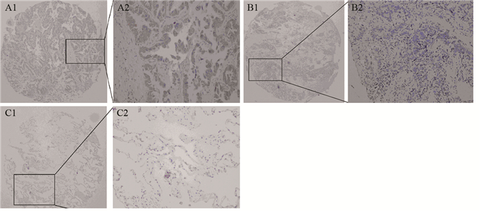
方法选取住院手术切除的肺腺癌组织及相应癌旁组织标本各80例,应用免疫组织化学法检测各组织中MAGE-As蛋白的表达,同时选取5例前列腺癌患者术后的睾丸组织作为阳性对照。
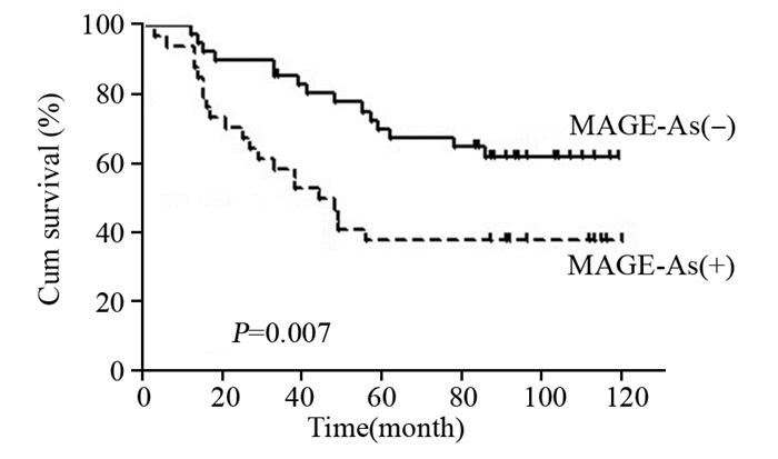
结果肺腺癌组织中MAGE-As蛋白的阳性表达率为46.25%(37/80),而相应的癌旁组织未发现MAGE-As蛋白的表达。MAGE-As蛋白的表达与肺癌患者的性别、年龄、临床分期、组织学分级、肿瘤大小、淋巴转移均无相关性(P > 0.05)。Log rank检验显示,MAGE-As蛋白表达阳性的肺腺癌患者的生存期均显著低于其表达阴性的患者(P=0.007)。多因素分析结果显示,MAGE-As表达、年龄和临床分期是肺腺癌患者较差预后的独立危险因素。
结论MAGE-As蛋白是肺腺癌的相关抗原。MAGE-As蛋白可能作为肺腺癌预后不良的指标。
-
关键词:
- 肺腺癌 /
- 黑色素瘤相关抗原-As /
- 组织芯片 /
- 预后
Abstract:ObjectiveTo investigate the expression of melanoma antigen (MAGE) -As in lung adenocarcinoma(LAC) tissues, and to explore their correlation with clinical biological indicators and prognostic factors.
MethodsWe collected cancerous and adjacent lung cancer tissue specimens from 80 patients with lung cancer who were surgically treated in the Fourth Hospital of Hebei Medical University. Testicular tissues (n=5) were collected from the patients with prostate cancer as positive control in the same time. Immunohistochemical staining was performed to assess the expression of MAGE-As in carcinoma and adjacent tissues.
ResultsMAGE-As protein were not expressed in adjacent tissues, the positive expression rate of MAGE-As protein in LAC tissues was 46.25%(37/80). No correlation was found between MAGE-As protein expression and age, gender, histological grade, clinical stage, tumor size, lymph node metastasis in LAC patients (P > 0.05). Log rank test showed that the survival time of LAC patients with positive MAGE-As expression was lower than that of negative ones (P=0.007). Multivariate analysis results showed that MAGE-As expression, age and clinical stage were independent risk factors of LAC.
ConclusionMAGE-As proteins are LAC-associated antigens, thus, they have potentially diagnostic and prognostic significance in clinical settings.
-
Key words:
- Lung adenocarcinoma /
- Melanoma antigen-As /
- Tissue microarray /
- Prognosis
-
0 引言
根据世界卫生组织国际癌症研究机构最新数据显示,2022年全球女性乳腺癌新发病例高达230万,位居全球癌症发病率第二位,仅次于肺癌[1]。国家癌症中心2022年发布的统计数据显示,2016年我国新发女性乳腺癌30.6万例,乳腺癌位居女性恶性肿瘤发病率第1位、死亡率第4位[2]。在乳腺癌的治疗中,多项临床研究显示中医药与放疗、化疗、靶向治疗、内分泌治疗等协同治疗,可以提高疗效、减轻不良反应,且疗效确切[3]。陈焕朝教授是湖北省名老中医,从事中医药治疗恶性肿瘤的临床工作四十余年,本研究通过数据挖掘分析陈焕朝教授治疗乳腺癌的中医处方,总结其用药规律,为中医药治疗乳腺癌提供参考。
1 资料与方法
1.1 病例来源
收集2022年8月—2023年11月陈焕朝教授在湖北省肿瘤医院门诊收治乳腺癌患者的处方数据。筛选治疗有效患者185例,录入首诊处方185首。
1.2 病例筛选标准
纳入标准:(1)西医诊断为乳腺癌,且有病理诊断;(2)基本信息完整,病历资料完善;(3)治疗期间按照医嘱服药3个月以上;(4)经治疗后症状确有改善,参考《中医病证诊断疗效标准》疗效评定有效者,录入首诊处方。
排除标准:排除乳腺癌合并其他恶性肿瘤的患者。
1.3 数据收集
收集患者一般资料,包括姓名、性别、年龄、处方数据,将相关数据录入 Excel 2019,建立数据库,所有录入信息由两位医师核对,确保数据的准确性。
1.4 数据标准化
参照2020版《中华人民共和国药典》[4]和《中药学》[5]对相关中药名称进行规范化,如“南方红豆杉”规范为“红豆杉”,“山萸肉”规范为“山茱萸”,“生黄芪” 规范为“黄芪”等,药物性味归经及功效亦参照《中华人民共和国药典》、《中药学》,整理后录入数据库。
1.5 统计学方法
通过Excel 2019建立药物数据库,并对药物频次、性味归经进行分析,得到频数分布雷达图。将185首处方导入SPSS Modeler 18.0建模后,选择Apriori算法,设置最低支持度为20%、最小置信度为80%,最大前项数为1,获得关联规则。运用SPSS Modeler 18.0对高频药物关联进行网络化展示。基于 SPSS 27.0对高频药物进行聚类分析,得出组方规律。
2 结果
2.1 药物频次
185例患者共涉及处方185首,中药180味,总频次为
3880 ,将中药频次由高到低进行排列,使用频次>40次的高频药物共29味,见表1。表 1 陈焕朝治疗乳腺癌处方高频药物(频次>40)Table 1 High-frequency drugs prescribed by Chen Huanchao for breast cancer treatment (frequency>40)序号 中药 频次 百分比(%) 序号 中药 频次 百分比(%) 序号 中药 频次 百分比(%) 1 黄芪 168 90.81 11 沙棘 101 54.59 21 莪术 58 31.35 2 茯苓 156 84.32 12 菝葜 98 52.97 22 鸡血藤 56 30.27 3 通关藤 153 82.70 13 白花蛇舌草 96 51.89 23 酸枣仁 50 27.03 4 桑黄 147 79.46 14 麸炒白术 93 50.27 24 补骨脂 48 25.95 5 夏枯草 132 71.35 15 郁金 82 44.32 25 杜仲叶 47 25.41 6 红景天 131 70.81 16 猪殃殃 73 39.46 26 柏子仁 45 24.32 7 蜈蚣 113 61.08 17 地黄 70 37.84 27 醋香附 45 24.32 8 远志 108 58.38 18 太子参 69 37.30 28 白英 42 22.70 9 天葵子 106 57.30 19 麸炒枳实 66 35.68 29 百药煎 41 22.16 10 红豆杉 101 54.59 20 姜厚朴 62 33.51 2.2 药物性味归经
180味中药药性以平、寒、温居多,频次分别为
1094 次、1055 次和896次,微温、微寒次之,分别为366次、363次;药味以甘、苦、辛为主,频次分别为2263 次、2014 次和1352 次;药物归经多属肝经、肺经、脾经和肾经,频次分别为1766 次、1615 次、1387 次和1361 次,其次为胃经1058 次、心经989次,见图1。2.3 药物功效分类
将185首处方中的中药按功效进行分类统计,使用频次由高到低依次是补虚药、清热药、利水渗湿药、化痰止咳平喘药、活血化瘀药、理气药等。排名前10位的药物功效见表2。
表 2 陈焕朝治疗乳腺癌处方常用药物功效归类(前10位)Table 2 Classification of the efficacy of commonly prescribed drugs used by Chen Huanchao in the treatment of breast cancer (top 10)排序 功效分类 味数 频次 频率(%) 1 补虚药 29 775 19.97 2 清热药 42 759 19.56 3 利水渗湿药 10 399 10.28 4 化痰止咳平喘药 17 366 9.43 5 活血化瘀药 14 274 7.06 6 理气药 12 192 4.9 7 平肝熄风药 8 178 4.59 8 止血药 4 176 4.54 9 安神药 7 170 4.38 10 消食药 8 167 4.30 2.4 药物组方关联规则分析
运用SPSS Modeler 18.0对185首处方进行关联规则分析,使用Apriori建模,设置最低支持度为20%、最小置信度为80%、最大前项数为1,获得120条关联规则,提升度表示前项出现对后项出现概率的影响,提升度>1且越高表明相关性越高。将所得关联规则按提升度降序排列并去重,前20位药物组合见表3。
表 3 185首治疗乳腺癌处方药物关联规则(支持度≥20%,置信度≥80%)Table 3 Association rules for the 185 breast cancer prescription drugs (support degree≥20%, confidence coefficient≥80%)后项 前项 支持度(%) 置信度(%) 提升度 姜厚朴 麸炒枳实 35.68 90.91 2.71 郁金 醋香附 24.32 95.56 2.16 麸炒白术 太子参 37.30 89.86 1.79 远志 酸枣仁 27.03 92.00 1.59 远志 柏子仁 24.32 91.11 1.58 菝葜 白英 22.70 83.33 1.57 天葵子 猪殃殃 39.46 82.19 1.43 蜈蚣 菝葜 52.97 83.67 1.37 蜈蚣 白花蛇舌草 51.89 83.33 1.36 蜈蚣 莪术 31.35 82.76 1.35 蜈蚣 红豆杉 54.59 80.20 1.31 夏枯草 莪术 31.35 87.93 1.24 夏枯草 天葵子 57.30 85.85 1.21 夏枯草 猪殃殃 39.46 84.93 1.20 夏枯草 郁金 44.32 84.15 1.19 夏枯草 蜈蚣 61.08 84.07 1.19 红景天 鸡血藤 30.27 83.93 1.19 夏枯草 菝葜 52.97 83.67 1.18 夏枯草 白英 22.70 83.33 1.18 夏枯草 红豆杉 54.59 83.17 1.17 桑黄 白英 22.70 92.86 1.17 夏枯草 白花蛇舌草 51.89 82.29 1.16 红景天 地黄 37.84 81.43 1.15 通关藤 猪殃殃 39.46 94.52 1.14 红景天 酸枣仁 27.03 80.00 1.13 红景天 柏子仁 24.32 80.00 1.13 桑黄 猪殃殃 39.46 89.04 1.12 桑黄 莪术 31.35 87.93 1.11 桑黄 菝葜 52.97 87.76 1.10 茯苓 麸炒白术 50.27 92.47 1.10 Note: The degree of improvement represents the effect of the occurrence of the preceding item on the probability of the following item appearing, and a degree of improvement greater than 1 indicates a high correlation. 运用SPSS Modeler 18.0对185首处方进行关联网络分析,药物节点间连线粗细和颜色深度代表药物间的关联程度,连线越粗、颜色越深表明药物间的关联程度越强。高频药物(使用频次>40)关联网络化展示见图2。
![]() 图 2 185首治疗乳腺癌处方高频药物关联网络化展示Figure 2 Network display of association among 185 high-frequency drugs prescribed for breast cancerThe thickness and color depth of the lines between drug nodes represent the degree of association between drugs. A thick line and a dark color indicate a strong degree of association between drugs.
图 2 185首治疗乳腺癌处方高频药物关联网络化展示Figure 2 Network display of association among 185 high-frequency drugs prescribed for breast cancerThe thickness and color depth of the lines between drug nodes represent the degree of association between drugs. A thick line and a dark color indicate a strong degree of association between drugs.2.5 药物系统聚类分析
将185首处方中使用频次>40次的高频药物共29味进行系统聚类分析,结合临床经验,大致可归为7类药物组合,见图3。
3 讨论
3.1 基于药物频数、性味归经、功效分析探讨用药规律
对药物频数进行分析发现,使用频率最高的前三味药分别是黄芪、茯苓、通关藤,黄芪味甘,性微温,归脾、肺经,为补气健脾要药。茯苓味甘、淡,性平,归心、肺、脾、肾经,可健脾、渗湿、利水消肿,黄芪、茯苓常配伍治疗脾气虚弱之证。通关藤味苦,性微寒,归肺经,具有止咳平喘、祛痰、清热解毒等作用。陈焕朝教授认为癌症的发生多因本虚,所谓“正气存内,邪不可干”、“邪之所凑,其气必虚”,而乳腺癌病位在肝,“见肝之病,知肝传脾,当先实脾”,故陈焕朝最常使用黄芪、茯苓补益脾气、培养中宫。在扶正培本之时,并用清热解毒祛痰之品以治标,标本兼治,相辅相成。现代药理研究表明,黄芪具有多种活性成分,其抗肿瘤作用成分研究主要集中于多糖类、皂苷类以及黄酮类成分,且对多种癌症,包括乳腺癌、肝癌、胃癌、结直肠癌、肺癌、卵巢癌等都具有明显的抑制作用[6]。黄芪可通过调节免疫功能、阻滞肿瘤细胞周期、促进肿瘤细胞凋亡与新血管生成、调节肿瘤细胞自噬、抑制炎性反应和逆转多药耐药性等多种途径发挥抗肿瘤作用[7-8]。茯苓主要活性成分以茯苓酸为代表的三萜类、以茯苓多糖为代表的多糖类等化合物,在抑制恶性肿瘤增殖与诱导凋亡、抑制侵袭转移、调节患者免疫功能及提高生活质量等方面表现出广泛的药理作用[9]。通关藤在体内外均具有抗肿瘤活性,抗肿瘤作用机制包括直接抗肿瘤作用,体现为抑制癌细胞增殖、调节肿瘤细胞血管生成、介导细胞凋亡、促进分化等,还包括对其他抗癌药物的增效减毒作用[10]。
对药物性味归经进行分析发现,药性以平为主,其次为寒性。平性药物寒热界限不太明显,药性平和,作用和缓。陈焕朝教授认为大多数乳腺癌患者病程较长,久病耗伤正气,脏腑虚损,应以平和药物和缓补之,否则体虚不受,或致壅滞经络,脏腑失调[11]。寒性药物可清热泻火、凉血解毒、清化热痰等,气滞、痰凝、血瘀郁久化热,患者常表现为心烦发热、乳房肿块红肿疼痛、便干尿黄等,适当应用寒性药物可有效缓解症状。药味以甘味为主,其次为苦味、辛味。甘能补、能和、能缓,具有补益和中、调和药性、缓急止痛的作用;苦能泄、能燥、能坚,具有清泄火热、燥湿、坚阴的作用;辛能散、能行,具有发散、行气血的作用[5]。陈焕朝教授认为乳腺癌患者多属本虚标实,宜扶正祛邪兼施,故甘苦辛同用,既可补益和中,又能清热燥湿、行气活血。归经分析结果显示药物主归肝经,其次为肺经、脾经、肾经和胃经。乳头属足厥阴肝经,乳房属足阳明胃经,女子以肝为先天,乳腺疾病与肝经密切相关。情志不遂、郁怒伤肝,肝失疏泄、经气郁滞,肝失条达横乘脾土,脾失健运则胃脏虚弱,脾胃运化失调则痰浊内生,痰凝气聚循经入乳,发为乳岩[12]。肝藏血、肾藏精,精血皆由水谷之精化生和充养,肝肾同源、藏泻互用,肝肾阴阳互滋互制,故陈焕朝教授治疗乳腺癌多从肝、脾、肾、胃论治。肝郁易化火、肝阳易上亢,辅以归肺经药物佐金平木,且乳腺癌发生肺转移风险较高,调理肺之宣发肃降功能尤为重要,因而陈焕朝教授处方中归肺经药物亦多见。
对药物功效进行分类,发现补益类药物居多,其次包括清热药、利水渗湿药、化痰止咳平喘药、活血化瘀药等,陈焕朝认为正气亏虚始终贯穿于乳腺癌发生、发展及转归的整个过程,邪实者,以气滞血瘀、湿聚痰积、毒踞为主,故应从虚、瘀、痰、毒论治[11]。正气亏虚多为脾肾虚衰、气血不足,予以健脾补肾、益气养血之品培本固元,瘀、痰、毒则以活血化瘀、化痰消积、清热解毒之品分消。乳腺癌发生发展的不同阶段,正气亏虚程度及夹杂有形实邪有所不同,临证过程中需仔细斟酌、辨证施治。
3.2 基于中药关联规则分析用药规律
药物关联规则中,姜厚朴-麸炒枳实置信度为90.91%,乳腺癌患者的最常见中医证型是肝郁气滞证[13],厚朴苦燥辛散,可消积导滞、燥湿消痰,既可除无形之湿满,又可消有形之实满。枳实辛行苦降,善破气行滞、化痰消积,二药合用可加强行气、化痰、消积之功。郁金-醋香附置信度为95.56%,郁金味辛,能行能散,既能入气分行气,又能入血分活血,香附主入肝经气分,芳香辛散,善散肝经之郁结,味苦疏泄可平肝气之横逆,为疏肝解郁、行气止痛之要药。麸炒白术-太子参置信度为89.86%,白术归脾、胃经,以补气、健脾、燥湿为主要作用,被誉为“补气健脾第一要药”,太子参补气健脾,兼以生津润肺,属补气药中清补之品,作用平和,陈焕朝教授认为,癌症为慢性疾病,补虚不宜太过,平补为主。
3.3 基于聚类分析探讨用药规律
通过高频药物聚类分析树状图可见,根据中医理论与临床实际,陈焕朝教授治疗乳腺癌常用药物聚拢较好的药物组合有两类。第一类组方为菝葜、白花蛇舌草、蜈蚣、天葵子、红豆杉。菝葜、蜈蚣、红豆杉通络散结、解毒散瘀,白花蛇舌草、天葵子清热解毒、消肿散结。此类药物均为陈焕朝教授治疗乳腺癌常用抗肿瘤中药,现代药理学研究均证实其具有抑制肿瘤作用。实验证明菝葜醇提物及其不同极性萃取部位对人乳腺癌细胞株MCF-7的增殖有抑制作用且呈现明显的量-效关系[14];白花蛇舌草总黄酮和蜈蚣提取液均可明显抑制乳腺癌MDA-MB-231细胞系增殖及促进细胞凋亡[15-16];天葵子抗肿瘤的有效部位为天葵子总生物碱,其抗肿瘤药效显著,药效机制可能与干预肿瘤炎性微环境,调控炎性因子表达以及相关蛋白的表达有关[17];红豆杉对PI3K/Akt/mTOR信号通路各靶点表达均有显著影响,对VEGF也有显著抑制作用[18]。陈焕朝教授认为在遵循辨证论治的基础上应用现代药理研究证实有抗肿瘤作用的中药,往往能收获更好的疗效。第二类组方为黄芪、茯苓、通关藤、桑黄、夏枯草、红景天。黄芪、茯苓、红景天健脾益气补中,脾胃为后天之本,气血生化之源,气血津液的化生均有赖于脾胃运化的水谷精微,李东垣《脾胃论·脾胃盛衰论》有云:“百病皆由脾胃衰而生也”,陈焕朝教授认为顾护脾胃应始终贯穿于乳腺癌治疗的整个过程。通关藤性微寒,有清热解毒、祛痰之功,桑黄亦性寒,有活血、化饮、软坚之效,夏枯草辛能散结、苦寒泄热,可治肝郁化火、痰火凝聚之证,在扶正的同时,应用清热解毒、化痰软坚之品,可谓攻补兼施,标本兼治,扶正而不留邪,祛邪而不伤正。
另外可得出五组药对,第一组为麸炒枳实、姜厚朴。第二组为郁金、醋香附。第三组为麸炒白术、太子参,与关联规则中得出的前三位药物组合一致。第四组药对为鸡血藤、补骨脂,晚期乳腺癌骨转移的发生率可高达75%[19],补骨脂温补脾肾,鸡血藤舒筋活络,两药相得益彰,可有效缓解骨转移导致的骨痛。第五组药对为酸枣仁、柏子仁,乳腺癌患者肝气郁结,肝郁化火,邪火扰动心神,神不安而不寐,酸枣仁、柏子仁养心益肝安神,常相须为用,为陈焕朝教授治疗失眠常用药对。
综上所述,本研究从客观数据资料出发,运用现代统计学方法,归纳总结了陈焕朝教授治疗乳腺癌的用药规律。陈教授认为乳腺癌基本病机为本虚标实,故以补虚类药物使用频率最高,顾护脾胃,培育先后天之本,始终贯穿于乳腺癌治疗的整个过程,热毒、痰湿、血瘀为标实,兼以清热解毒、化痰消积、活血化瘀等治法,攻补兼施,可取得良好疗效。本研究仅对陈焕朝教授治疗乳腺癌用药经验进行初步探析,以期为中医药治疗乳腺癌提供思路。
-
表 1 肺腺癌组织中MAGE-As和VEGF蛋白的阳性表达与患者临床病理特征之间的关系
Table 1 Correlation of positive MAGE-As VEGF expression with clinicopathological features of LAC patients

表 2 影响肺腺癌患者10年生存率预后因素的单因素及多因素分析
Table 2 Univariate and multivariate analyses of prognostic factors for 10-year survival of LAC patients

-
[1] Siegel RL, Miller KD, Jemal A. Cancer statistics, 2015[J]. CA Cancer J Ciln, 2015, 65(1): 5-29. doi: 10.3322/caac.21254
[2] Chen W, Zheng R, Baade PD, et al. Cancer statistics in China, 2015[J]. CA Cancer J Clin, 2016, 66(2): 115-32. doi: 10.3322/caac.21338
[3] Salmaninejad A, Zamani MR, Pourvahedi M, et al. Cancer/Testis antigens: expression, regulation, tumor invasion and use in immunotherapy of cancers[J]. Immuol Invest, 2016, 45(7): 619-40. doi: 10.1080/08820139.2016.1197241
[4] Van der Bruggen P, Traversari C, Chomez P, et al. A gene encoding an antigen recognized by cytolytic T lymphocytes on a human melanoma[J]. Science, 1991, 254 (5038): 1643-7. doi: 10.1126/science.1840703
[5] Wang D, Wang J, Ding N, et al. MAGE-A1 promotes melanoma proliferation and migration through C-JUN activation[J]. Biochem Biophys Res Commun, 2016, 473(4): 959-65. doi: 10.1016/j.bbrc.2016.03.161
[6] Hou SY, Sang MX, Geng CZ, et al. Expressions of MAGE-A9 and MAGE-A11 in breast cancer and their expression mechanism[J]. Arch Med Res, 2014, 45(1): 44-51. doi: 10.1016/j.arcmed.2013.10.005
[7] Liu SH, Sang M, Xu Y, et al. Expression of MAGE-A1, -A9, -A11 in laryngeal squamous cell carcinoma and their prognostic significance: a retrospective clinical study[J]. Acta Otolaryngol, 2016, 136(5): 506-13. doi: 10.3109/00016489.2015.1126856
[8] Mengus C, Schultz-Thater E, Coulot J, et al. MAGE-A10 cancer/testis antigen is highly expressed in high-grade non-muscle-invasive bladder carcinomas[J]. Int J Cancer, 2013, 132(10): 2459-63. doi: 10.1002/ijc.27914
[9] 谷丽娜, 桑梅香, 刘飞, 等.黑素瘤相关抗原-A9和-A11在食管鳞状细胞癌组织中的表达及其临床意义[J].中国肿瘤生物治疗杂志, 2015, 22(5): 630-6. doi: 10.3872/j.issn.1007-385X.2015.05.014 Gu LN, Sang MX, Liu F, et al. expression and clinical significance of melanoma antigen-a9 and a11 in esophageal squamous cell caricinoma tissues[J]. Zhongguo Zhong Liu Sheng Wu Zhi Liao Za Zhi, 2015, 22(5): 630-6. doi: 10.3872/j.issn.1007-385X.2015.05.014
[10] Guo L, Sang M, Liu Q, et al. The expression and clinical significance of melanoma-associated antigen-A1, -A3 and -A11 in glioma[J]. Oncol Lett, 2013, 6(1): 55-62.
[11] 刘维华, 桑梅香, 单保恩.黑色素瘤相关抗原-A:食管癌免疫治疗新靶点[J].肿瘤防治研究, 2015, 42(6): 622-6. http://www.zlfzyj.com/CN/abstract/abstract8525.shtml Liu WH, Sang MX, Shan BE. MAGE-A: New Target of Immunotherapy for Esophageal Cancer[J]. Zhong Liu Fang Zhi Yan Jiu, 2015, 42(6): 622-6. http://www.zlfzyj.com/CN/abstract/abstract8525.shtml
[12] Brahmer JR. Harnessing the immune system for the treatment of non-small-cell-lung cancer[J]. J Clin Oncol, 2013, 31(8): 1021-8. doi: 10.1200/JCO.2012.45.8703
[13] Pujol JL, Vansteenkiste JF, De Pas TM, et al. Safety and Immunogenicity of MAGE-A3 Cancer Immunotherapeutic with or without Adjuvant Chemotherapy in Patients with Resected Stage ⅠB to Ⅲ MAGE-A3-Positive Non-Small-Cell Lung Cancer[J]. J Thorac Oncol, 2015, 10(10): 1458-67. doi: 10.1097/JTO.0000000000000653
[14] Vansteenkiste J, Zielinski M, Linder A, et al. Adjuvant MAGE-A3 immunotherapy in resected non-small-cell lung cancer: phaseⅡ randomized study results[J]. J Clin Oncol, 2013, 31(1): 2396-403. doi: 10.1200/JCO.2012.43.7103
[15] Karimi S, Mohammadi F, Porabdollah M, et al. Characterization of melanoma-associated antigen-a genes family differential expression in non-small-cell lung cancers[J]. Clin Lung Cancer, 2012, 13(3): 214-9. doi: 10.1016/j.cllc.2011.09.007
[16] Vansteenkiste JF, Cho BC, Vanakesa T, et al. Efficacy of the MAGE-A3 cancer immunotherapeutic as adjuvant therapy in patients with resected MAGE-A3-positive non-small-cell lung cancer (MAGRIT): a randomised, double-blind, placebo-controlled, phase 3 trial[J]. Lancet Oncol, 2016, 17(6): 822-35. doi: 10.1016/S1470-2045(16)00099-1
[17] Sienel W, Varwerk C, Linder A, et al. Melanoma associated antigen (MAGE)-A3 expression in Stages Ⅰ and Ⅱ non-small cell lung cancer: results of a multi-center study[J]. Eur J Cardiothorac Surg, 2004, 25 (1): 131-4. doi: 10.1016/j.ejcts.2003.09.015
[18] Peikert T, Specks U, Farver C, et al. Melanoma antigen A4 is expressed in non-small cell lung cancers and promotes apoptosis[J]. Cancer Res, 2006, 66(9): 4693-700. doi: 10.1158/0008-5472.CAN-05-3327
[19] Karimi S, Mohammadi F, Porabdollah M, et al. Characterization of melanoma-associated antigen-a genes family differential expression in non-small-cell lung cancers[J]. Clin Lung Cancer, 2012, 13(3): 214-9. doi: 10.1016/j.cllc.2011.09.007
[20] Zhang S, Zhai X, Wang G, et al. High expression of MAGE-A9 in tumor and stromal cells of non-small cell lung cancer was correlated with patient poor survival[J]. Int J Clin Exp Pathol, 2015, 8(1): 541-50. https://www.ncbi.nlm.nih.gov/pmc/articles/PMC4348844/
[21] Zhai X, Xu L, Zhang S, et al. High expression levels of MAGE-A9 are correlated with unfavorable survival in lung adenocarcinoma[J]. Oncotarget, 2016, 7(4): 4871-81. doi: 10.18632/oncotarget.v7i4
[22] Adair SJ, Hogan KT. Treatment of ovarian cancer cell lines with 5-aza-2'-deoxycytidine upregulates the expression of cancer-testis antigens and class Ⅰ major histocompatibility complex-encoded molecules[J]. Cancer Immunol Immunother, 2009, 58(4): 589-601. doi: 10.1007/s00262-008-0582-6
[23] Liu XL, Sun N, Dong YN, et al. Anticancer effects of adenovirus-mediated calreticulin and melanoma-associated antigen 3 expression on non-small cell lung cancer cells[J]. Int Immunopharmacol, 2015, 25(2): 416-24. doi: 10.1016/j.intimp.2015.02.017
[24] Jin J, Liu BZ, Wu ZM. Evaluation of melanoma antigen gene A3 expression in drug resistance of epidermal growth factor receptor-tyrosine kinase inhibitors in advanced non-small cell lung cancer treatment[J]. J Cancer Res Ther, 2015, 11(8): 271-4. doi: 10.4103/0973-1482.170549


 下载:
下载:


