
Expression of IRSp53 in Epithelial Ovarian Carcinoma and Its Prognostic Significance
-
摘要:目的
探讨胰岛素受体酪氨酸激酶底物p53蛋白(IRSp53)在上皮性卵巢癌(epithelial ovarian cancer, EOC)中的表达及其对患者预后的影响。
方法采用SP免疫组织化学法检测76例不同病理类型EOC组织中IRSp53蛋白的表达,分析其表达与患者的临床病理特征和病理分级的关系,探讨IRSp53对EOC患者预后的影响。
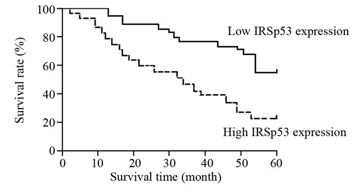
结果IRSp53蛋白的表达与FIGO分期(Z=-3.526, P < 0.05)、组织病理分级(Z=-2.471, P < 0.05)、病理学类型(Z=-2.305, P < 0.05)、淋巴结转移(Z=1.124, P < 0.05)和CA125水平(Z=1.462, P < 0.05)显著相关。IRSp53的表达强度与病理分级之间呈显著正相关性(r=0.779, P < 0.05)。IRSp53蛋白低水平表达患者的5年累积生存率显著高于IRSp53蛋白高水平表达者(χ2=5.018, P < 0.05)。
结论IRSp53蛋白可能是EOC发生发展过程中的关键调控因子,其过度表达导致患者的预后更差。
Abstract:ObjectiveTo analyse the IRSp53 expression and its prognostic significance in epithelial ovarian carcinoma tissues.
MethodsThe expressions of IRSp53 in 76 cases of epithelial ovarian carcinoma were detected by SP immunohistochemical technique. The relationship between the IRSp53 expression and the clinical pathological characteristics and the pathological grading of the patients was analyzed. The prognostic significance of IRSp53 in epithelial ovarian carcinoma was discussed.
ResultsThe expression of IRSp53 was significantly correlated with FIGO stage (Z=-3.526, P < 0.05), histopathological grading (Z=-2.471, P < 0.05), pathologic type (Z=-2.305, P < 0.05), lymph node metastasis (Z=1.124, P < 0.05) and CA125 level (Z=1.462, P < 0.05). There was a positive correlation between the expression intensity of IRSp53 and pathological grade (r=0.779, P < 0.05). The 5-year cumulative survival rate of the patients with low IRSp53 expression was significantly higher than that with high IRSp53 expression (χ2 =5.018, P < 0.05).
ConclusionIRSp53 plays an important role in the development of epithelial ovarian carcinoma, and the over-expression of IRSp53 may lead to poor prognosis.
-
Key words:
- Epithelial ovarian cancer /
- IRSp53 /
- Prognosis
-
-
![]()
图 2 IRSp53蛋白表达水平与EOC患者预后关系
Figure 2 Relationship between IRSp53 expression and prognosis of EOC patients
表 1 IRSp53在EOC组织中的表达与临床病理特征关系
Table 1 Relationship between IRSp53 and clinical characteristics in EOC

 下载: 导出CSV
表 2 IRSp53在EOC组织中的表达及其与病理分级的关系 (n=76)
Table 2 Relationship between IRSp53 and pathological grading in EOC (n=76)
 下载: 导出CSV
-
[1] Chambers SK. Role of CSF-1 in progression of epithelial ovarian cancer[J]. Future Oncol, 2016, 5(9): 1429-40. https://www.researchgate.net/publication/38079817_Role_of_CSF-1_in_progression_of_epithelial_ovarian_cancer
[2] Nassir M, Guan J, Luketina H, et al. The role of HE4 for prediction of recurrence in epithelial ovarian cancer patients-results from the OVCAD study[J]. Tumour Biol, 2016, 37(3): 3009-16. doi: 10.1007/s13277-015-4031-9
[3] Zhang Z, Zhou B, Gao Q, et al. A polymorphism at miRNA-122-binding site in the IL-1α 3'UTR is associated with risk of epithelial ovarian cancer[J]. Fam Cancer, 2014, 13(4): 595-601. doi: 10.1007/s10689-014-9739-y
[4] Colombo PE, Fabbro M, Theillet C, et al. Sensitivity and resistance to treatment in the primary management of epithelial ovarian cancer[J]. Crit Rev Oncol Hematol, 2014, 89(2): 207-16. doi: 10.1016/j.critrevonc.2013.08.017
[5] Chieng CK, Say YH. Cellular prion protein contributes to LS 174T colon cancer cell carcinogenesis by increasing invasiveness and resistance against doxorubicin-induced apoptosis[J]. Tumour Biol, 2015, 36(10): 8107-20. doi: 10.1007/s13277-015-3530-z
[6] Girault A, Privé A, Trinh NT, et al. Identification of KvLQT1 K+ channels as new regulators of non-small cell lung cancer cell proliferation and migration[J]. Int J Oncol, 2014, 44(3): 838-48. https://www.spandidos-publications.com/ijo/44/3/838?text=fulltext
[7] Chung W, Choi SY, Lee E, et al. Social deficits in IRSp53 mutant mice improved by NMDAR and mGluR5 suppression[J]. Nat Neurosci, 2015, 18(3): 435-43. doi: 10.1038/nn.3927
[8] Thomas A, Mariani-Floderer C, López-Huertas MR, et al. Involvement of the Rac1-IRSp53-Wave2-Arp2/3 Signaling Pathway in HIV-1 Gag Particle Release in CD4 T Cells[J]. J Virol, 2015, 89(16): 8162-81. doi: 10.1128/JVI.00469-15
[9] Karimi-Zarchi M, Mortazavizadeh SM, Bashardust N, et al. The Clinicopathologic Characteristics and 5-year Survival Rate of Epithelial Ovarian Cancer in Yazd, Iran[J]. Electron Physician, 2016, 8(1): 1399-406. http://europepmc.org/articles/PMC4623803
[10] Koh SC, Razvi K, Chan YH, et al. The association with age, human tissue kallikreins 6 and 10 and hemostatic markers for survival outcome from epithelial ovarian cancer[J]. Arch Gynecol Obstet, 2011, 284(1): 183-90. doi: 10.1007/s00404-010-1605-z
[11] Lu Y, Huang S, Li P, et al. Prognostic evaluation of preoperative serum C-reactive protein concentration in patients with epithelial ovarian cancer[J]. Exp Ther Med, 2015, 9(5): 2003-7. https://www.spandidos-publications.com/etm/9/5/2003?text=fulltext
[12] López-Contreras L, Hernández-Ramírez VI, Flores-García Y. Src and PI3 K inhibitors affect the virulence factors of Entamoeba histolytica[J]. Parasitology, 2013, 140(2): 202-9. doi: 10.1017/S0031182012001540
[13] Chen CJ, Shih CH, Chang YJ, et al. SH2B1 and IRSp53 proteins promote the formation of dendrites and dendritic branches[J]. J Biol Chem, 2015, 290(10): 6010-21. doi: 10.1074/jbc.M114.603795
[14] Chen H, Wu X, Pan ZK, et al. Integrity of SOS1/EPS8/ABI1 tri-complex determines ovarian cancer metastasis[J]. Cancer Res, 2010, 70(23): 9979-90. doi: 10.1158/0008-5472.CAN-10-2394
[15] Kast DJ, Yang C, DisanzaA, et al. Mechanism of IRSp53 inhibition and combinatorial activation by Cdc42 and downstream effectors[J]. Nat Struct Mol Biol, 2014, 21(4): 413-22. doi: 10.1038/nsmb.2781
[16] Oikawa T, Okamura H, Dietrich F, et al. IRSp53 mediates podosome formation via VASP in NIH-Src cells[J]. PLoS One, 2013, 8(3): e60528. doi: 10.1371/journal.pone.0060528
计量
- 文章访问数: 1151
- HTML全文浏览量: 333
- PDF下载量: 271

