
Relationship of Macrophage Migration Inhibitory Factor Expression with Clinicopathologic Features and Prognosis of Cardiac Carcinoma Patients
-
摘要:目的
探讨巨噬细胞移动抑制因子(macrophage migration inhibitory factor, MIF)在贲门癌中的表达及其与患者临床病理特征和预后的关系。
方法选取滨州医学院附属医院胸外科2008年1月—2009年12月经胸手术切除的贲门癌石蜡标本73例,采用免疫组织化学法检测贲门癌组织中MIF的表达水平,根据免疫反应强弱将其分为高表达组和低表达组,分析MIF表达水平与患者临床病理特征的关系,Kaplan-Meier生存曲线比较MIF高表达组和低表达组患者的生存期,Cox回归模型分析MIF的表达水平与临床病理特征及患者预后的关系。
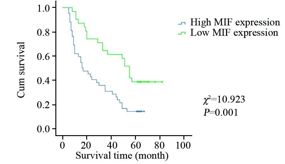
结果MIF在贲门癌组织中高表达42例,高表达率为57.5%。MIF表达水平与患者的年龄、性别、肿瘤分化程度、淋巴结转移和TNM分期无关(P>0.05),与肿瘤的浸润深度有关(P=0.025)。MIF高表达组和低表达组的生存期用Kaplan-Meier方法评估,并用Log rank检验进行比较,两组之间生存率差异有统计学意义(P=0.001)。多因素Cox回归模型分析表明,淋巴结转移(P=0.025)、MIF表达水平(P=0.001)是影响贲门癌预后的独立危险因素。
结论MIF的表达异常可能在贲门癌的浸润及预后中有重要作用,对患者预后的判断有一定指导意义。
-
关键词:
- 贲门癌 /
- 巨噬细胞移动抑制因子 /
- 临床病理特征 /
- 预后
Abstract:ObjectiveTo investigate the expression of macrophage migration inhibitory factor (MIF) in cardiac carcinoma and its correlation with clinicopathologic features and prognosis.
MethodsThe paraffin specimens from 73 patients with cardiac carcinoma who underwent thoracotomy were collected from the Affiliated Hospital of Binzhou Medical University from 2008-01-03 to 2009-12-31. The expressions of MIF in 73 specimens of cardiac carcinoma were assessed by immunohistochemistry. Based on the levels of MIF immunoreactivities, the 73 specimens were divided into high MIF expression group and low MIF expression group, and the correlations of MIF expression with clinicopathologic features was analyzed. Kaplan-Meier survival curves were used to compare the survival between high MIF expression group and low MIF expression group. The correlations of MIF expression with clinicopathologic features and the prognosis of cardiac carcinoma patients were analyzed by Cox regression model.
ResultsThe high MIF expression rate was 57.5%. There was no significant correlation between the levels of MIF expression and age, gender, tumor differentiation, lymph node metastasis, TNM stages(P>0.05). However, the high MIF expression was strongly correlated with the depth of invasion(P=0.025). The survival rates were significantly different between the two groups(P=0.001). The multivariate Cox regression model analysis showed that lymph node metastasis(P=0.025) and the levels of MIF expression(P=0.001) were the independent prognostic risk factors for cardiac carcinoma.
ConclusionThe abnormal of MIF expression in cardiac carci noma may play an important role in tumor infiltration and prognosis. MIF could be a valuable prognostic factor for the patients with cardiac carcinoma.
-
Key words:
- Cardiac carcinoma /
- MIF /
- Clinicopathologic features /
- Prognosis
-
0 引言
淋巴管浸润和淋巴结转移是结直肠癌患者预后较差的重要原因之一,新生淋巴管是肿瘤淋巴道转移的基础和前提[1-2]。DNA结合分化抑制蛋白1(inhibitors of DNA binding-1,Id-1)是近年来研究较热门的致癌基因,能促进细胞增殖,诱导肿瘤血管内皮生成,促进肿瘤的生长及侵袭,其表达的缺失、沉默与肺癌、胃癌、胰腺癌、宫颈癌等多种恶性肿瘤发生、发展及预后密切相关[3-6]。MMP-9能够降解细胞外各型胶原、层粘连蛋白、蛋白聚糖等,诱导肿瘤细胞突破基底膜致其浸润,参与肿瘤脉管形成[7-8]。D-240特异性表达于肿瘤微淋巴管细胞,在血管内皮细胞中几无表达,因而D-240能准确监测癌组织内的微淋巴管密度(lymphatic microvessel density,LMVD),反映肿瘤淋巴管新生状况。本实验通过对结直肠癌组织中Id-1、MMP-9和LMVD行免疫组织化学分析,探讨三者与结直肠癌临床相关病理的联系及其相关性,为探索结直肠癌演进、淋巴结转移的机制及抗淋巴管浸润治疗提供科学依据。
1 资料与方法
1.1 资料来源
收集2013年5月1日—2014年5月31日河北北方学院附属第一医院血管腺体外科手术切除的原发性结直肠癌组织50例、癌旁正常组织50例,癌组织取自癌灶中心处,另取标本残端切缘经病理证实的正常结直肠组织,术前均未行新辅助放化疗等针对性治疗。其中男32例、女18例,年龄33~71(51±2.9)岁。
1.2 免疫组织化学染色方法及试剂
兔抗人单克隆浓缩型Id-1抗体、鼠抗人浓缩型MMP-9单克隆抗体均购于英国Abcam公司;D-240抗体购于美国Abnova公司。10%甲醛溶液固定部分标本,石蜡包埋,切片厚度为4 µm,苏木精-伊红(hematoxylin-esin,HE)染色,80℃烤箱烤片,乙醇脱水,灭活内源性过氧化物酶,枸橼酸缓冲液高温高压水化修复;PBS冲洗3次,加一抗,4℃过夜;PBS洗3次,加二抗。室温下DAB显色,苏木精轻度对比染色,并用1%的盐酸乙醇分化5 min、流水冲洗5 min、梯度乙醇脱水(80%、85%、90%、95%、100%、100%)及二甲苯透明,干燥,最后树胶封片。阴性对照为PBS代替一抗。
1.3 Id-1、MMP-9阳性判定标准
Id-1和MMP-9均以细胞质中出现棕黄色颗粒为阳性表达,根据着色细胞占视野细胞总数的百分比及着色细胞染色强弱评分≤5%为0分,5%~25%为1分,>25%~50%时为2分,>50%~75%时为3分,>75%时为4分;阴性:0分;弱(+):1分;中(++ ):2分;强(+++):3分。两者相乘,0分为(-),1~4分为(+),5~8分为(++),9~12分为( +++),(+)~(+++)均视为阳性。
1.4 微淋巴管密度检测
应用D-240抗体标记染色于肿瘤微淋巴管内皮细胞的胞质,以孤立的单个棕黄色内皮细胞或内皮细胞簇作为微淋巴管并计数;先在40倍光学显微镜下选出4个脉管最密集的区域(即富集热点区),后换200倍光学显微镜下将着色的内皮细胞区计为观察区,计数取其平均值即为LMVD值。
1.5 统计学方法
运用SPSS17.0统计软件统计并分析数据,用百分率表示计数资料,运用χ2检验表示Id-1和MMP-9的表达与临床病理参数关系,用(x±s)表示计量资料,运用t检验表示LMVD值与临床病理参数的关系,相关性分析采用Spearmen检验,设α=0.05为检验水准。
2 结果
2.1 Id-1和MMP-9在结直肠癌中的表达及LMVD值
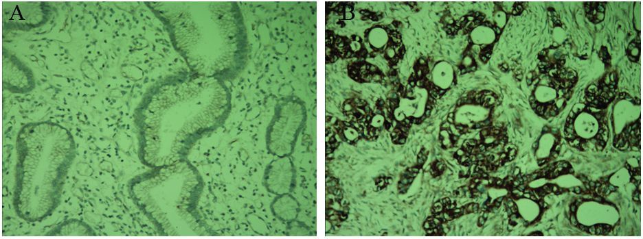
Id-1在结直肠癌组织中的阳性表达72.00%(36/50)明显高于癌旁组织24.00%(12/50),两者比较差异有统计学意义(χ2=23.431,P=0.000); MMP-9在结直肠癌组织中的阳性表达为78%(39/50),明显高于癌旁组织28.00%(14/50),两者比较差异有统计学意义(χ2=18.944,P=0.000);结直肠癌组织中LMVD表达(15.18±2.16)明显高于癌旁组织(5.24±1.09),两者比较差异有统计学意义(t=25.051,P=0.000),见图 1,表 1。
![]() 图 1 免疫组织化学法检测Id-1、MMP-9在结直肠癌癌旁与癌组织中的表达和LMVDFigure 1 Expression of Id-1,MMP-9 and LMVD in colorectal normal adjacent and colorectal tissues detected by IHC methodA: positive expression of Id-1 in colorectal normal adjacent tissues; B: positive expression of Id-1 in colorectal carcinoma tissues; C : positive expression of MMP-9 in colorectal normal adjacent tissues; D: positive expression of MMP-9 in colorectal carcinoma tissues; E: positive expression of LMVD markd by D-240 in colorectal carcinoma tissues (IHC ×200)表 1 Id-1、MMP-9在结直肠癌癌旁与癌组织中的表达和LMVDTable 1 The expressions of Id-1,MMP-9 and LMVD in colorectal normal adjacent tissues and colorectal carcinoma tissues
图 1 免疫组织化学法检测Id-1、MMP-9在结直肠癌癌旁与癌组织中的表达和LMVDFigure 1 Expression of Id-1,MMP-9 and LMVD in colorectal normal adjacent and colorectal tissues detected by IHC methodA: positive expression of Id-1 in colorectal normal adjacent tissues; B: positive expression of Id-1 in colorectal carcinoma tissues; C : positive expression of MMP-9 in colorectal normal adjacent tissues; D: positive expression of MMP-9 in colorectal carcinoma tissues; E: positive expression of LMVD markd by D-240 in colorectal carcinoma tissues (IHC ×200)表 1 Id-1、MMP-9在结直肠癌癌旁与癌组织中的表达和LMVDTable 1 The expressions of Id-1,MMP-9 and LMVD in colorectal normal adjacent tissues and colorectal carcinoma tissues
2.2 Id-1和MMP-9蛋白的表达水平及LMVD值与临床病理参数间的关系
Id-1、MMP-9表达水平和LMVD均与肿瘤浆膜浸润、TNM分期、淋巴结转移、肝转移、脉管浸润、CEA(+)等相关(均P<0.01),见表 2。
表 2 Id-1和MMP-9在结直肠癌组织中的表达及LMVD与临床病理因素的关系Table 2 The relationship between the expression of Id-1,MMP-9,LMVD value and clinical pathological parameters in colorectal carcinoma tissues
2.3 Id-1和MMP-9在结直肠腺癌组织中的表达与LMVD的关联
结直肠癌中Id-1阳性者36例,其LMVD值(18.26±3.14);Id-1阴性者14例,其LMVD值(10.09±3.03),两者比较差异有统计学意义(t=18.511,P=0.000);结直肠癌中MMP-9阳性者39例,其LMVD值(19.11±3.20);MMP-9阴性者11例,其LMVD值(9.21±3.05),两者比较差异有统计学意义(t=21.770,P=0.000)。
2.4 Id-1和MMP-9在结直肠癌中表达的相关性
Id-1和MMP-9在结直肠癌中的表达呈明显的正相关关系(r=0.429,P<0.01),见表 3。
表 3 结直肠癌组织Id-1和MMP-9表达的相关性Table 3 The correlation between the expression of Id-1 and MMP-9 in colorectal carcinoma tissues
3 讨论
Id-1系螺旋-环-螺旋(helix-loop-helix,HLH)转录因子家族重要成员,在正常组织中呈低表达或不表达状态,而在恶性肿瘤及体外培养的肿瘤细胞系中均呈高表达趋势,且与肿瘤的恶性度及患者的预后相关[9]。大多数HLH结构存在一个碱性HLH(bHLH)因子与其紧密相邻,两者相互结合形成异质二聚体,其碱性因子所处区域与目标DNA结合,诱导启动子中的E-盒样结构的靶基因转录并整合成所谓的“E-box”DNA序列,从而调控细胞增殖分化。而Id-1无碱性结构域,与bHLH因子结合生成的二聚体无转录功能,因而无法与目标DNA序列结合,从而抑制细胞分化增殖[10-11]。Cheung等[12]研究报道Id-1表达上调能激活Raf-1及MAPK激酶信号转导途径、抑制细胞凋亡、参与肿瘤发生发展。Lee等[13]研究发现,Id-1还可通过磷酸化抑制Rb途径并阻断p16(INK4a)表达,从而促进G1/S期细胞增殖。
肿瘤的浸润转移是多因素、多步骤参与的复杂过程,其中细胞基质的降解和破坏是肿瘤淋巴道和血行转移的关键步骤。MMP-9作为一种特异性极高血管内皮分裂原,对肿瘤脉管形成及肿瘤恶性生物学行为均有非常重要的影响。Redondo-Munoz等[14]研究显示MMP-9能通过降解细胞外基质使癌细胞呈阿米巴运动方式穿过基质膜缺损处迁移至内皮细胞进入淋巴管腔内,促进淋巴道转移;魏礼清、黄榕权等[15-16]对胰腺癌和浸润性乳腺癌组织研究显示MMP-9能通过裂解细胞外基质,诱导淋巴管内皮细胞迁移从而促进微淋巴管形成,且由于新生淋巴管内皮较薄,基底膜不完整,因而癌细胞易于进入淋巴管道,致淋巴结转移。
本研究显示Id-1和MMP-9在结直肠癌组织中表达明显高于癌旁组织,且与肿瘤的浆膜浸润、TNM分期、淋巴结转移、肝转移及脉管浸润等密切相关,支持Id-1和MMP-9促进肿瘤恶性生物学进展的理论,这与胡斌等[17]研究一致。 LMVD是评价肿瘤微淋巴管生成重要指标,能够准确反映肿瘤微淋巴管生成状况,本实验表明结直肠癌组织中LMVD表达明显高于癌旁组织,同时Id-1和MMP-9在结直肠癌中的表达呈正相关性,且与LMVD亦存在明显的正相关性,进一步说明Id-1和MMP-9在参与结直肠癌变发生发展过程中具有协同作用,Id-1的高异常表达能激活MMP-9,释放促脉管生成的自分泌信号,诱导形成新生的脉管内皮细胞,从而促进肿瘤的淋巴道转移,这与徐健、Georgiadou等[18-19]研究基本一致。且有研究[20]表明 Id-1能通过上调VEGF的表达诱导肿瘤微血管的生成,促进结直肠癌血行转移。Fong等[21]对伴转移的乳腺癌细胞的研究发现抑制Id-1的表达,MT1-MMP表达水平也随之下调,癌细胞的侵袭能力亦明显降低,表明Id-1通过诱导MMP蛋白的表达,诱导微淋巴管形成,发挥促进肿瘤淋巴道转移的作用。
总之,Id-1的异常表达参与结直肠癌的发生演进,并与淋巴结转移等恶性生物学行为密切相关,但本研究仅限于分子蛋白水平,未能从基因水平及其致癌的具体机制进行深入研究,这亦是本课题今后的努力方向。
-
![]()
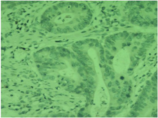
图 1 贲门癌组织中MIF的表达 (IHC×400)
Figure 1 Expression of MIF in cardiac carcinoma tissues (IHC×400)
![]()
图 2 胃正常组织中MIF的阴性表达 (IHC×400)
Figure 2 Negative MIF expression in gastric mucosae tissues (IHC×400)
![]()
图 3 MIF高表达和低表达与贲门癌患者5年生存率的关系
Figure 3 Relationship between high and low expression of MIF and 5-year survival rates of cardiac carcinoma patients
表 1 MIF表达与贲门癌患者临床病理特征的关系
Table 1 Relationship between MIF expression and clinicopathological features of cardiac carcinoma patients

 下载: 导出CSV
表 2 贲门癌患者多因素Cox模型分析结果
Table 2 Multivariate analysis of cardiac carcinoma patients by Cox regression
 下载: 导出CSV
-
[1] Morris KT, Nofchissey RA, Pinchuk IV, et al. Chronic macrophage migration inhibitory factor exposure induces mesenchymal epithelial transition and promotes gastric and colon cancers[J]. PLoS One, 2014, 9(6): e98656. doi: 10.1371/journal.pone.0098656
[1] Morris KT, Nofchissey RA, Pinchuk IV, et al. Chronic macrophage migration inhibitory factor exposure induces mesenchymal epithelial transition and promotes gastric and colon cancers[J]. PLoS One, 2014, 9(6): e98656. [2] Babu SN, Chetal G, Kumar S. Macrophage migration inhibitory factor: a potential marker for cancer diagnosis and therapy[J]. Asian Pac J Cancer Prev, 2012, 13(5): 1737-44. [2] Babu SN, Chetal G, Kumar S. Macrophage migration inhibitory factor: a potential marker for cancer diagnosis and therapy[J].Asian Pac J Cancer Prev, 2012, 13(5): 1737-44. doi: 10.7314/APJCP.2012.13.5.1737
[3] Zheng YX, Yang M, Rong TT, et al. CD74 and marcophage migration inhibitory factor as therapeutic targets in gastric cancer[J]. World J Gastroenterol, 2012, 18(18): 2253-61. [3] Zheng YX, Yang M, Rong TT, et al. CD74 and marcophage migration inhibitory factor as therapeutic targets in gastric cancer[J]. World J Gastroenterol, 2012, 18(18): 2253-61. doi: 10.3748/wjg.v18.i18.2253
[4] Simpson KD, Templeton DJ, Cross JV. Macrophage Migration Inhibitory Factor promotes tumor growth and metastasis by inducing Myeloid Derived Suppressor Cells in the tumor microenvironment[J]. J Immunol, 2012, 189(12): 5533-40. doi: 10.4049/jimmunol.1201161
[4] Simpson KD, Templeton DJ, Cross JV. Macrophage Migration Inhibitory Factor promotes tumor growth and metastasis by inducing Myeloid Derived Suppressor Cells in the tumor microenvironment[J]. J Immunol, 2012, 189(12): 5533-40. [5] Gámez-Pozo A, Sánchez-Navarro I, Calvo E, et al. PTRF/cavin-1 and MIF proteins are identified as non-small cell lung cancer biomarkers by label-free Proteomics[J]. PLoS One, 2012, 7(3): e33752. doi: 10.1371/journal.pone.0033752
[5] Gámez-Pozo A, Sánchez-Navarro I, Calvo E, et al. PTRF/cavin-1 and MIF proteins are identified as non-small cell lung cancer biomarkers by label-free Proteomics[J]. PLoS One, 2012, 7(3): e33752. [6] Zhang L,Ye SB, Ma G, et al. The expressions of MIF and CXCR4 protein in tumor microenvironment are adverse prognostic factors in patients with esophageal squamous cell carcinoma[J]. J Transl Med, 2013, 11: 60. doi: 10.1186/1479-5876-11-60
[6] Zhang L,Ye SB, Ma G, et al. The expressions of MIF and CXCR4 protein in tumor microenvironment are adverse prognostic factors in patients with esophageal squamous cell carcinoma[J]. J Transl Med, 2013, 11: 60. [7] Li H, Zang J, Wang P, et al. Gastric cancer susceptibility in gastric cancer relatives: attributable risks of Macrophage migration inhibitory factor promoter polymorphism and Helicobacter pylori[J]. Cytokine, 2012, 60(2): 346-51. [7] Li H, Zang J, Wang P, et al. Gastric cancer susceptibility in gastric cancer relatives: attributable risks of Macrophage migration inhibitory factor promoter polymorphism and Helicobacter pylori[J]. Cytokine, 2012, 60(2): 346-51. doi: 10.1016/j.cyto.2012.07.015
[8] Li J, Mo HY, Xiong G, et al. Tumor microenvironment macrophage inhibitory factor directs the accumulation of interleukin-17-producing tumor-infiltrating lymphocytes and predicts favorable survival in nasopharyngeal carcinoma patients[J]. J Biol Chem, 2012, 287(42): 35484-95. doi: 10.1074/jbc.M112.367532
[8] Li J, Mo HY, Xiong G, et al. Tumor microenvironment macrophage inhibitory factor directs the accumulation of interleukin-17- producing tumor-infiltrating lymphocytes and predicts favorable survival in nasopharyngeal carcinoma patients[J]. J Biol Chem, 20 12, 287(42): 35484-95. [9] 蔡滕, 吴苏稼, 樊根涛. 等. 巨噬细胞移动抑制因子在骨肉瘤中 的表达及其与预后的关系[J]. 临床肿瘤学杂志, 2013, 18(2): 12 3-5. [Cai T, Wu SJ, Fan GT, et al. Expression of macrophage migration inhibitory factor in osteosarcoma and its correlation with prognosis[J]. Lin Chuang Zhong Liu Xue Za Zhi, 2013, 18 (2): 123-5.] [9] 蔡滕, 吴苏稼, 樊根涛. 等. 巨噬细胞移动抑制因子在骨肉瘤中的表达及其与预后的关系[J]. 临床肿瘤学杂志, 2013, 18(2): 123-5. http://www.cnki.com.cn/Article/CJFDTOTAL-LCZL201302008.htm Cai T, Wu SJ, Fan GT, et al. Expression of macrophage migration inhibitory factor in osteosarcoma and its correlation with prognosis[J]. Lin Chuang Zhong Liu Xue Za Zhi, 2013, 18(2): 123-5. http://www.cnki.com.cn/Article/CJFDTOTAL-LCZL201302008.htm
[10] 夏娟, 李冬, 郑伟萍, 等. 炎症与肿瘤的关系研究进展[J]. 国际检验医学杂志, 2013, 34(1): 63-4. http://www.cnki.com.cn/Article/CJFDTOTAL-GWSQ201301033.htm Xia J, Li D, Zheng WP, et al. Research progress on relationship between inflammation and tumor[J]. Guo Ji Jian Yan Yi Xue Za Zhi, 2013, 34(1): 63-4. http://www.cnki.com.cn/Article/CJFDTOTAL-GWSQ201301033.htm
[10] 夏娟, 李冬, 郑伟萍, 等. 炎症与肿瘤的关系研究进展[J]. 国际 检验医学杂志, 2013, 34(1): 63-4. [Xia J, Li D, Zheng WP, et al. Research progress on relationship between inflammation and tumor[J]. Guo Ji Jian Yan Yi Xue Za Zhi, 2013, 34(1): 63-4.] [11] 赵德利, 陈万青, 于婷婷, 等. 贲门癌发病危险因素的病例对照 研究[J] 中华肿瘤杂志, 2011, 33(10): 775-8. [Zhao DL, Chen WQ, Ding TT, et al. A population-based matched case-control study on the risk factors of gastric cardia cancer[J]. Zhonghua Zhong Liu Za Zhi, 2011, 33(10): 775-8.] [11] 赵德利, 陈万青, 于婷婷, 等. 贲门癌发病危险因素的病例对照研究[J]. 中华肿瘤杂志, 2011, 33(10): 775-8. http://www.cqvip.com/qk/93685x/201110/39798275.html Zhao DL, Chen WQ, Ding TT, et al. A population-based matched case-control study on the risk factors of gastric cardia cancer[J]. Zhonghua Zhong Liu Za Zhi, 2011, 33(10): 775-8. http://www.cqvip.com/qk/93685x/201110/39798275.html
[12] 欧玉荣, 康敏, 周蕾, 等. 胃癌中幽门螺旋杆菌L型感染与MIF、 MMP9、VEGF表达的关系[J]. 南方医科大学学报, 2014, 34(2): 18 0-7. [Ou YR, Kang M, Zhou L, et al. Infection with L-form of Helicobacter pylori and expressions of MIF, MMP9 and VEGF in gastric carcinoma[J]. Nan Fang Yi Ke Da Xue Xue Bao, 2014, 34 (2): 180-7.] [12] 欧玉荣, 康敏, 周蕾, 等. 胃癌中幽门螺旋杆菌L型感染与MIF、MMP9、VEGF表达的关系[J]. 南方医科大学学报, 2014, 34(2): 180-7. http://www.cnki.com.cn/Article/CJFDTOTAL-DYJD201402008.htm Ou YR, Kang M, Zhou L, et al. Infection with L-form of Helicobacter pylori and expressions of MIF, MMP9 and VEGF in gastric carcinoma[J]. Nan Fang Yi Ke Da Xue Xue Bao, 2014, 34(2): 180-7. http://www.cnki.com.cn/Article/CJFDTOTAL-DYJD201402008.htm
[13] Arenberg D, Luckhardt TR, Carskadon S, et al. Macrophage migration inhibitory factor promotes tumor growth in the context of lung injury and repair[J]. Am J Respir Crit Care Med, 2010, 18 2(8): 1030-7. [13] Arenberg D, Luckhardt TR, Carskadon S, et al. Macrophage migration inhibitory factor promotes tumor growth in the context of lung injury and repair[J]. Am J Respir Crit Care Med, 2010, 182(8): 1030-7. doi: 10.1164/rccm.201001-0120OC
[14] 徐慧鲜, 吴礼浩, 马伟钦, 等. 靶向MIF的siRNA对大肠癌细胞增殖的影响[J]. 中华全科医学, 2013, 11(3): 334-5. http://www.cnki.com.cn/Article/CJFDTOTAL-SYQY201303003.htm Xu HX, Wu LH, Ma WQ, et al. The Effect of the Small Interfering Rnas for Macrophage Migration Inhibitory Factor on the Proliferation of Colorectal Cancer Cell Line[J]. Zhonghua Quan Ke Yi Xue, 2013, 11(3): 334-5. http://www.cnki.com.cn/Article/CJFDTOTAL-SYQY201303003.htm
[14] 徐慧鲜, 吴礼浩, 马伟钦, 等. 靶向MIF的siRNA对大肠癌细胞 增殖的影响[J]. 中华全科医学, 2013, 11(3): 334-5. [Xu HX, Wu LH, Ma WQ, et al. The Effect of the Small Interfering Rnas for Macrophage Migration Inhibitory Factor on the Proliferation of Colorectal Cancer Cell Line[J]. Zhonghua Quan Ke Yi Xue, 2013, 11 (3): 334-5.] [15] 张洪典, 唐鹏, 段晓峰, 等. 贲门癌淋巴结外软组织转移的临床病理特征及预后分析[J]. 中华外科杂志, 2013, 51(10): 882-6. Zhang HD, Tang P, Duan XF, et al. Analysis of the clinicopathologic characters and prognostic impact of extranodal metastasis in gastric cardia patients[J]. Zhonghua Wai Ke Za Zhi, 2013, 51(10): 882-6.
[15] 张洪典, 唐鹏, 段晓峰, 等. 贲门癌淋巴结外软组织转移的 临床病理特征及预后分析[J]. 中华外科杂志, 2013, 51(10): 88 2-6. [Zhang HD, Tang P, Duan XF, et al. Analysis of the clinicopathologic characters and prognostic impact of extranodal metastasis in gastric cardia patients[J]. Zhonghua Wai Ke Za Zhi, 20 13, 51(10): 882-6.] [16] 丁学伟, 王晓娜, 薛强, 等. 贲门癌的临床病理特征分析[J]. 中 华肿瘤防治杂志, 2010, 17(10): 770-2. [Ding XW, Wang XN, Xue Q, et al. Clinicomathological characteristics of gastric cardia cancer[J]. Zhonghua Zhong Liu Fang Zhi Za Zhi, 2010, 17(10): 77 0-2.] [16] 丁学伟, 王晓娜, 薛强, 等. 贲门癌的临床病理特征分析[J]. 中华肿瘤防治杂志, 2010, 17(10): 770-2. http://www.cnki.com.cn/Article/CJFDTOTAL-QLZL201010018.htm Ding XW, Wang XN, Xue Q, et al. Clinicomathological characteristics of gastric cardia cancer[J]. Zhonghua Zhong Liu Fang Zhi Za Zhi, 2010, 17(10): 770-2. http://www.cnki.com.cn/Article/CJFDTOTAL-QLZL201010018.htm
[17] Hosokawa Y, Kinoshita T, Konishi M, et al. Clinicopathological features and prognostic factors of adenocarcinoma of the esophagogastric junction according to Siewert classification: experiences at a single institution in Japan[J]. Ann Surg Oncol, 20 12, 19(2): 677-83. [17] Hosokawa Y, Kinoshita T, Konishi M, et al. Clinicopathological features and prognostic factors of adenocarcinoma of the esophagogastric junction according to Siewert classification: experiences at a single institution in Japan[J]. Ann Surg Oncol, 2012, 19(2): 677-83. doi: 10.1245/s10434-011-1983-x

计量
- 文章访问数: 1252
- HTML全文浏览量: 347
- PDF下载量: 222

