
Expressions of MACC1 and c-Met Proteins in Lung Adenocarcinoma and Their Correlation with Postoperative Recurrence
-
摘要:目的
检测MACC1、c-Met蛋白在肺腺癌组织中的表达,及其在患者术后复发中的预测价值。
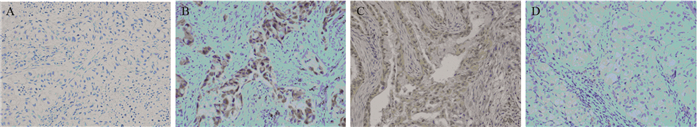
方法收集完全性切除的肺腺癌患者术后病理标本102例和癌旁组织57例,采用免疫组织化学技术检测MACC1和c-Met蛋白在肺腺癌和癌旁组织中的表达,对患者行定期随访,分析MACC1和c-Met蛋白的表达与患者术后复发的关系。
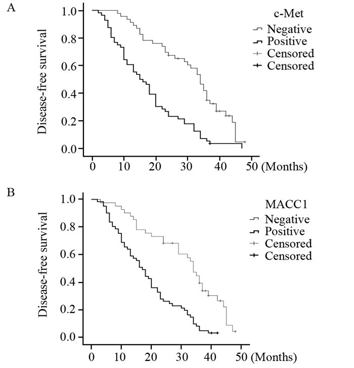
结果MACC1和c-Met在癌组织中的阳性表达率分别为59.80%、54.90%,明显高于癌旁组织中的7.01%、10.53%,差异有统计学意义(P<0.05)。MACC1、c-Met蛋白的表达与性别年龄无关,与肿瘤T分期、淋巴结转移、病理TNM分期有关(P<0.05)。MACC1与c-Met的表达之间呈正相关(r=0.262, P=0.008)。MACC1阳性组2年内复发率为73.77%,明显高于阴性组的31.71%(χ2=17.686, P<0.001)。c-Met阳性组2年内复发率为76.79%,高于阴性组的34.78%(χ2=18.272, P<0.001)。Cox回归多因素提示MACC1和c-Met蛋白的表达、术后病理分期、肿瘤T分期及淋巴结转移情况是腺癌患者复发转移的危险因素。
结论MACC1和c-Met蛋白在肺腺癌组织中表达与肿瘤T分期、淋巴结转移、病理分期有关,影响肺腺癌患者术后无瘤生存期,是复发转移的危险因素。
-
关键词:
- 结肠癌转移相关基因1 /
- c-Met /
- 肺腺癌 /
- 复发
Abstract:ObjectiveTo detect the expressions of metastasis associated in colon cancer 1(MACC1) and c-Met proteins in patients with lung adenocarcinoma and analyze the suitability of these protein expressions to predict postoperative recurrence.
MethodsThere were 102 lung adenocarcinoma patients who underwent a completely surgical excision from July 2011 to July 2013. The expression of MACC1 and c-Met proteins in lung adenocarcinoma tissues and normal lung tissues were detected by immunohistochemistry. We had the regular follow-up for all patients. The correlation between protein expressions and postoperative recurrence was analyzed.
ResultsIn 102 cases of lung adenocarcinoma tissues and 57 cases of normal lung tissues, the positive MACC1 protein expression rate was higher in tumor tissues than in normal lung tissues(59.80% vs. 7.01%, P<0.05). The positive c-Met protein expression rate was higher in tumor tissues than in normal tissues (54.90% vs. 10.53% , P<0.05). The gender and age were not found to be correlated with MACC1 or c-Met protein expressions, while significant association were shown between the expressions of MACC1, c-Met proteins and pathological T stage, lymph node metastasis, TNM stage(P<0.05). The MACC1 and c-Met protein expressions were positively correlated(r=0.262, P=0.008). The relapse rate within two years with MACC1 positive expression was 73.77% and higher than 31.71% with negative expression(χ2=17.686, P<0.001). There were different 2-year relapse rates between positive and negative expressions of c-Met protein(76.79% vs. 34.78%, χ2=18.272, P<0.001). Cox regression analysis showed that MACC1 expression, c-Met expression, postoperative pathological stage, T stage and lymph node metastasis were the prognostic factors of the recurrence and metastasis of lung adenocarcinoma.
ConclusionThe expression levels of MACC1 and c-Met protein are associated with T stage, lymph node metastasis and pathological stage, and are the prognostic factors of the recurrence and metastasis of lung adenocarcinoma.
-
Key words:
- Metastasis associated in colon cancer1(MACC1) /
- c-Met /
- Lung adenocarcinoma /
- Recurrence
-
0 引言
宫颈癌是威胁全球女性生命健康的第二大恶性肿瘤,发病率仅次于乳腺癌,全世界每年约有50万的新发宫颈癌病例,其中80%来自发展中国家,且发病率和病死率一直居高不下,宫颈癌的治疗一直是国内外研究的重点领域[1-2]。miRNA是一类单链内源性非编码小分子RNA,能够通过与靶mRNA特异性的碱基配对引起靶mRNA的降解或者抑制其翻译,从而对基因进行转录后的表达调控。目前已经证实miRNA在生物发育和细胞分化中发挥重要作用,越来越多的研究表明,miRNA参与多种细胞生物过程与癌症的发生、发展和转移密切相关[3-5]。
自噬是真核细胞的一种非细胞凋亡性程序性死亡,又被称为Ⅱ型程序性细胞死亡。当细胞受到不良刺激如营养缺乏、内质网应激、氧化应激、细胞器损伤以及细胞死亡的情况下都可以激活自噬信号通路,在多种人类肿瘤中存在细胞自噬活性改变,自噬活性降低促进肿瘤的发生和进展[6-8]。自噬在神经退行性疾病、感染性疾病及肿瘤发生发展过程中的作用逐渐被揭示,细胞自噬防止坏死引起的无菌性炎性反应和巨噬细胞浸润,不仅可以限制肿瘤细胞生长,同时也可以抑制肿瘤侵袭转移[9-11],GFP-LC3目前被认为是自噬体特异性标签,是一种检测自噬发生常用的实验技术手段。
为了深入了解宫颈癌发生发展的分子机制和寻找新的治疗靶点,我们通过对miR-216b在宫颈癌细胞中作用的研究,为寻求宫颈癌特异性靶向诊断和治疗提供新的思路。
1 材料与方法
1.1 材料
人宫颈癌细胞系HeLa购自中国科学院上海细胞库;RPMI 1640细胞培养液及胎牛血清购自美国Life Technologies公司;GFP-LC3质粒由南京大学医学院惠赠。雷帕霉素、Beclin-1、LC-3B、GAPDH等抗体购自美国Santa Cruz公司;胰蛋白酶与胎牛血清购自美国Gibco公司;miR-216b引物、mimic和inhibitor及内参U6均购自广州锐博生物科技有限公司,转染所用阳离子脂质体LipofectamineTM 3000及Opti-MEM购自美国Invitrogen公司。
1.2 方法
1.2.1 细胞自噬率检测
将HeLa细胞接种到24孔板内,培养过夜,使细胞密度达到70%~80%。通过LipofectamineTM 3000转染GFP-LC3质粒体,6 h后换完全培养液继续培养24 h,之后将miR-216b mimic或inhibitor分别转染48 h,阳性对照组给予5 μmol/L雷帕霉素处理24 h。将处理后的细胞用PBS清洗3次,用4%多聚甲醛室温固定15 min,PBS清洗3次,荧光显微镜下观察,统计GFP-LC3的阳性自噬点数,统计对照组和处理组单个细胞中GFP-LC3荧光点数,每组至少统计60个细胞。
1.2.2 Western blot法检测自噬相关蛋白表达
将HeLa细胞均匀接种于60 mm培养皿中,药物处理后用预冷的PBS洗3遍,加入含有100 mmol/L PMSF的裂解液(RIPA)裂解细胞,冰上放置30 min充分裂解,4℃条件下12 000 r/min离心15 min,取上清液后经BCA法测定蛋白浓度。依次进行质量分数10%的SDS-PAGE凝胶电泳,使用PVDF转膜,5%脱脂奶粉封闭2 h后,4℃封闭一抗过夜,TBST清洗3次(每次5 min)后加入相应二抗,室温下孵育60 min,TBST洗3次后进行显影,实验重复三次,采用计算机软件ImageJ分析灰度值并记录。
1.2.3 miR-216b表达水平的检测
6孔板内接种HeLa细胞各6×10⁵个。待细胞完全贴壁后用miR-216b inhibitor转染48 h,TRIzol试剂裂解细胞,按照试剂说明书提取基因组总RNA,取<0.5 μg RNA为模板进行反转录合成cDNA。按照说明书混合PCR反应体系(SYBRGreen),每管分别加入1 μl cDNA产物,仪器设置条件为:95℃ 10 min;95℃ 15 s,60℃ 1 min,共计40个循环。
1.2.4 报告基因质粒的构建与检测
构建pmirGLO荧光素酶质粒报告系统(美国Promega公司)分析miRNA与预测靶序列的结合,该质粒含有两个不同荧光素酶基因,一个为插入Beclin-1 3’UTR序列的荧光素酶基因表达载体(野生型),另一个基因为靶结合位点突变的载体质粒(突变型)。将两种质粒分别与miR-216b mimic及其阴性对照共转染HeLa细胞,每组设5个复孔,培养48 h后进行荧光素酶相对活性检测,双荧光素酶报告实验(Dual Luciferase report assay)(美国Promega公司)上机检测。
1.3 统计学方法
采用SPSS19.0统计学软件进行统计分析,经K-S检验,本研究各检测指标的数据资料呈正态分布,以均数±标准差表示,组间均数经Levene检验方差齐。采用均衡分组单因素干预多水平实验设计,处理因素作用总体差异比较均采用单因素方差分析,两两比较分别采用LSD-t检验和SNK-q检验。P<0.05被认为差异具有统计学意义。
2 结果
2.1 miR-216b抑制宫颈癌细胞自噬水平
HeLa细胞转染GFP-LC3后,再转染miR-216b mimic,以雷帕霉素作为阳性对照,在荧光显微镜下观察并对带有GFP-LC3的自噬点阳性细胞数和总细胞数进行统计。相对于对照组和雷帕霉素组的细胞,miR-216b能够明显降低细胞自噬体的数量,见图 1。Western blot检测结果显示,自噬标记蛋白Beclin1表达量减少,作为自噬发生指标的LC3-Ⅱ/LC3-I比值也同样表现出下降;miR-216b能够降低HeLa细胞自噬蛋白的表达水平,见图 2。综上结果表明,miR-216b能够抑制HeLa细胞的自噬水平。
![]() 图 1 荧光显微镜观察经miR-216b转染或雷帕霉素处理后HeLa细胞中GFP-LC3自噬点的分布变化Figure 1 Distribution of GFP-LC3 autophagy points in HeLa cells after transfection of miR-216b or rapamycin treatment observed by fluorescence microscopyA: the autophagy positive cells in normal control group,miR-216b mimic group and rapamycin group were observed by fluorescence microscope. Bright green fluorescent spots represented autophagy positive cells and the pictures below were for low power lens to corresponding groups;B: Autophagy positive cell rates in normal control group,miR-216b mimic group and rapamycin group (*: P<0.05)
图 1 荧光显微镜观察经miR-216b转染或雷帕霉素处理后HeLa细胞中GFP-LC3自噬点的分布变化Figure 1 Distribution of GFP-LC3 autophagy points in HeLa cells after transfection of miR-216b or rapamycin treatment observed by fluorescence microscopyA: the autophagy positive cells in normal control group,miR-216b mimic group and rapamycin group were observed by fluorescence microscope. Bright green fluorescent spots represented autophagy positive cells and the pictures below were for low power lens to corresponding groups;B: Autophagy positive cell rates in normal control group,miR-216b mimic group and rapamycin group (*: P<0.05)![]() 图 2 Western blot检测miR-216b抑制细胞自噬水平Figure 2 Cell autophagy inhibited by miR-216b detected by Western blotA: the expression of Beclin-1,LC3-Ⅰ and LC3-Ⅱ in normal control group,miR-216b mimic group and rapamycin group detected by Western blot; B: the ratio of LC3-Ⅱ/LC3-Ⅰ in normal control group,miR-216b mimic group and rapamycin group (*: P<0.05)
图 2 Western blot检测miR-216b抑制细胞自噬水平Figure 2 Cell autophagy inhibited by miR-216b detected by Western blotA: the expression of Beclin-1,LC3-Ⅰ and LC3-Ⅱ in normal control group,miR-216b mimic group and rapamycin group detected by Western blot; B: the ratio of LC3-Ⅱ/LC3-Ⅰ in normal control group,miR-216b mimic group and rapamycin group (*: P<0.05)2.2 抑制miR-216b的表达能够促进宫颈癌细胞的自噬水平
转染miR-216b抑制剂的细胞中,miR-216b表达量出现明显下降,下降近80%。同时,其自噬点阳性细胞数明显增多,见图 3。通过Western blot检测发现,自噬相关蛋白Beclin-1和LC3-Ⅱ的表达均表现出增加,见图 4。结果表明,在HeLa细胞中,抑制miR-216b的表达能够促进自噬水平的升高。
![]() 图 3 荧光显微镜观察经miR-216b抑制剂转染处理后HeLa中GFP-LC3自噬点的分布变化Figure 3 Distribution of GFP-LC3 autophagy points in HeLa cells after transfection with miR-216b inhibitor observed by fluorescence microscopyA: the expression of miR-216b in anti-NC group and anti-miR-216b group after transfection with miR-216b inhibitors; B: the positive rate of autophagy points in anti-NC group and anti-miR-216b group after transfection with miR-216b inhibitor; C: the autophagy positive cells in anti-NC group and anti-miR-216b group observed by fluorescence microscope after transfection with miR-216b inhibitor. Bright green fluorescent spots represented autophagy positive cells
图 3 荧光显微镜观察经miR-216b抑制剂转染处理后HeLa中GFP-LC3自噬点的分布变化Figure 3 Distribution of GFP-LC3 autophagy points in HeLa cells after transfection with miR-216b inhibitor observed by fluorescence microscopyA: the expression of miR-216b in anti-NC group and anti-miR-216b group after transfection with miR-216b inhibitors; B: the positive rate of autophagy points in anti-NC group and anti-miR-216b group after transfection with miR-216b inhibitor; C: the autophagy positive cells in anti-NC group and anti-miR-216b group observed by fluorescence microscope after transfection with miR-216b inhibitor. Bright green fluorescent spots represented autophagy positive cells![]() 图 4 Western blot检测发现抑制miR-216b表达能够促进细胞自噬水平Figure 4 Inhibiting miR-216b expression could promote cells autophagy level observed by Western blotA: the expression of Beclin-1,LC3-Ⅰ,LC3-Ⅱ in anti-NC group and anti-miR-216b mimic group after transfection with miR-216b inhibitors detected by Western blot; B: the ratio of LC3-Ⅱ/LC3-Ⅰ in NC-anti group and anti-miR-216b mimic group after transfection with miR-216b inhibitors (*: P<0.05)
图 4 Western blot检测发现抑制miR-216b表达能够促进细胞自噬水平Figure 4 Inhibiting miR-216b expression could promote cells autophagy level observed by Western blotA: the expression of Beclin-1,LC3-Ⅰ,LC3-Ⅱ in anti-NC group and anti-miR-216b mimic group after transfection with miR-216b inhibitors detected by Western blot; B: the ratio of LC3-Ⅱ/LC3-Ⅰ in NC-anti group and anti-miR-216b mimic group after transfection with miR-216b inhibitors (*: P<0.05)2.3 miR-216b能够靶向结合Beclin-1
查找数据库发现,miR-216b能够与Beclin-1的3’UTR区域中的579-585段结合,见图 5结果显示。两种质粒分别与miR-216b mimic及其阴性对照共转染HeLa细胞,48 h后将细胞裂解,通过荧光素值检测发现,转染Beclin-1 3’UTR质粒载体(野生组)和miR-216b组,其荧光素值相对于对照组明显下降,而Beclin-1 3’UTR突变位点组(突变组)荧光素值相对于对照组,未出现明显变化,见图 6。 结果表明,miR-216b能够通过靶结合Beclin-1中的3‘UTR区域中的579~585段发挥其抑制作用。
![]() 图 5 数据库分析miR-216b能够靶结合Beclin-1 3’UTR序列Figure 5 miR-216b could targetedly binding Beclin-1 3'UTR sequence analyzed by database
图 5 数据库分析miR-216b能够靶结合Beclin-1 3’UTR序列Figure 5 miR-216b could targetedly binding Beclin-1 3'UTR sequence analyzed by database![]() 图 6 荧光素酶报告基因检测miR-216b能够靶结合Beclin-1 3’UTRFigure 6 miR-216b could targetedly binding Beclin-1 3'UTR detected by Luciferase
图 6 荧光素酶报告基因检测miR-216b能够靶结合Beclin-1 3’UTRFigure 6 miR-216b could targetedly binding Beclin-1 3'UTR detected by Luciferase3 讨论
MicroRNAs是真核生物细胞中一类长度约为17~24个核苷酸的内源性非编码小RNA。它本身不编码蛋白质,主要通过负性调控靶基因的表达发挥作用。近年来研究表明,某些miRNA的差异性表达与宫颈癌的发生、进展有密切关系,可以作为宫颈癌的诊断及预后的指标。宫颈癌中miRNA表达的失调与肿瘤的增殖、凋亡、侵袭和转移有重要关系[3-5]。
近年来miRNA在调节肿瘤自噬水平方面受到广泛关注,在维持细胞内环境稳定方面起着重要作用。本实验中,通过Western blot和GFP-LC3 shRNA转染细胞等检测方法,首次发现miR-216b能够显著降低HeLa细胞自噬水平,而施加miR-216b抑制剂后可明显提高自噬水平。本实验所选取的自噬相关蛋白-微管相关蛋白1轻链3(microtubule-associated protein 1 light chain 3,LC3)是目前研究较多的自噬标志性物质,其定位于前自噬泡和自噬泡膜表面,参与自噬体的形成,被认为是自噬特异性形成指标;Beclin-1是哺乳动物的自噬调控基因,是自噬发生的触发器,其表达的上调可诱导自噬的形成,可通过促进细胞凋亡诱导细胞自噬而抑制肿瘤的发展过程[12]。两种自噬指示蛋白在转染miR-216b mimic的作用下,均出现表达量降低。然而,转染miR-216b抑制剂后,自噬水平出现了上调,这与前述结果吻合,说明miR-216b的确能够抑制宫颈癌细胞的自噬水平。为进一步研究miR-216b在自噬过程中的功能,本研究通过荧光素酶报告基因的检测发现miR-216b能够靶结合Beclin-1的3’UTR区域,进而发挥其抑制作用。
Beclin-1是酵母菌ATG6在哺乳类动物的同源基因,是第一个被发现参与自噬过程的基因,其编码的Beclin-1蛋白可调控自噬前体的形成,引导相关蛋白定位于自噬体膜[13]。体外研究发现,Beclin-1缺失的小鼠自发和病毒诱导的肿瘤发生率增加[14-15],说明Beclin-1在抑制肿瘤发生方面有重要作用。本实验发现miR-216b能够负调控Beclin-1的表达来影响宫颈癌细胞自噬水平。
越来越多的研究发现机体细胞自噬活性变化对肿瘤的发生发展及对抗肿瘤药物的药效影响具有双重作用。研究发现自噬相关蛋白在宫颈癌中表达受抑制,下调自噬的形成,促进肿瘤的发生和发展[16],另外,也有研究发现,当自噬相关蛋白过表达后,宫颈癌细胞的生长会受到明显抑制[17],因此,通过调控自噬形成有可能成为未来宫颈癌治疗的策略之一,而本实验所研究的miR-216b的功能就是能够抑制宫颈癌细胞自噬的形成。宫颈癌是威胁女性健康的恶性肿瘤,目前,仍无有效的治疗方式。而自噬是维持细胞稳态的重要通路,在疾病中的作用逐步得到认同。本研究首次表明了miR-216b在宫颈癌自噬发生过程中的机制,为治疗宫颈癌提供了理论基础,也为开发类似药物提供了新的思路。
-
![]()
图 2 c-Met(A)和MACC1(B)蛋白阳性表达与阴性表达患者的无病生存期曲线
Figure 2 Disease-free survival curves of patients with positive and negative c-Met(A) and MACC1(B) expression
表 1 c-Met和MACC1蛋白表达与肺腺癌患者临床及病理特点的关系
Table 1 Clinical and pathological characteristics of lung adenocarcinoma patients with c-Met and MACC1 overexpression

 下载: 导出CSV
表 2 Cox多因素分析肺腺癌患者复发的危险因素
Table 2 Cox multivariate analysis of risk factors for patients with lung adenocarcinoma recurrence
 下载: 导出CSV
-
[1] 李学祥, 王慜杰, 高佳, 等. 肺肿瘤患者临床病理特征回顾性分析[J].中华肿瘤防治杂志, 2012, 19(2): 130-3. http://www.cnki.com.cn/Article/CJFDTOTAL-QLZL201202015.htm Li XX, Wang MJ, Gao J, et al. Retrospective analysis of clinical pathology characteristic in patients with lung neoplasms[J].Zhonghua Zhong Liu Fang Zhi Za Zhi, 2012, 19(2): 130-3. http://www.cnki.com.cn/Article/CJFDTOTAL-QLZL201202015.htm
[1] Li XX, Wang MJ, Gao J, et al. Retrospective analysis of clinical pathology characteristic in patients with lung neoplasms[J]. Zhonghua Zhong Liu Fang Zhi Za Zhi, 2012, 19(2): 130-3. [李学 祥, 王慜杰, 高佳, 等. 肺肿瘤患者临床病理特征回顾性分析[J]. 中华肿瘤防治杂志, 2012, 19(2): 130-3.] [2] Goya T, Asamura H, Yoshimura H, et al. Prognosis of 6644 resected non-small cell lung cancers in Japan:a Japanese lung cancer registry study[J]. Lung Cancer, 2005, 50(2): 227-34. doi: 10.1016/j.lungcan.2005.05.021
[2] Goya T, Asamura H, Yoshimura H, et al. Prognosis of 6644 resected non-small cell lung cancers in Japan:a Japanese lung cancer registry study[J]. Lung Cancer, 2005, 50(2): 227-34. [3] Stein U. MACC1-a novel target for solid cancers[J]. Expert Opin Ther Targets, 2013, 17(9): 1039-52. http://cn.bing.com/academic/profile?id=2083029824&encoded=0&v=paper_preview&mkt=zh-cn
[3] Stein U. MACC1-a novel target for solid cancers[J]. Expert Opin Ther Targets, 2013, 17(9): 1039-52. [4] Gumustekin M, Kargi A, Bulut G, et al. HGF/c-Met overexpression, but not met mution, correlates with progression of non-small cell lung cancer[J]. Pathol Oncol Res, 2012, 18(2): 209-18. [4] Gumustekin M, Kargi A, Bulut G, et al. HGF/c-Met overexpression, but not met mution, correlates with progression of non-small cell lung cancer[J]. Pathol Oncol Res, 2012, 18(2): 209-18. doi: 10.1007/s12253-011-9430-7
[5] Qiu J, Huang P, Liu Q, et al. Identification of MACC1 as a novel prognostic marker in hepatocellular carcinoma[J]. J Transl Med, 2011, 9: 166. doi: 10.1186/1479-5876-9-166
[5] Qiu J, Huang P, Liu Q, et al. Identification of MACC1 as a novel prognostic marker in hepatocellular carcinoma[J]. J Transl Med, 20 11, 9: 166. [6] Fan X, Zhang X, Wang H, et al. Reevaluation of survival and prognostic factors in pathologic stage adenocarcinoma by the new 20 09 TNM classification[J]. Tumor Biol, 2014, 35(6): 5905-10. [6] Fan X, Zhang X, Wang H, et al. Reevaluation of survival and prognostic factors in pathologic stage adenocarcinoma by the new 2009 TNM classification[J]. Tumor Biol, 2014, 35(6): 5905-10. doi: 10.1007/s13277-014-1781-8
[7] Shimokawa H, Uramoto H, Onitsuka T, et al. Overexpression of MACC1 mRNA in lung adenocarcinoma is associated with postoperative recurrence[J]. J Thorac Cardiovasc Surg, 2011, 14 1(4): 895-8. [7] Shimokawa H, Uramoto H, Onitsuka T, et al. Overexpression of MACC1 mRNA in lung adenocarcinoma is associated with postoperative recurrence[J]. J Thorac Cardiovasc Surg, 2011, 141(4): 895-8. doi: 10.1016/j.jtcvs.2010.09.044
[8] Park S, Choi YL, Sung CO, et al. High MET copy number and MET overexpression: poor outcome in non-small cell lung cancer patients[J]. Histol Histopathol, 2012, 27(2): 197-207. http://cn.bing.com/academic/profile?id=285988926&encoded=0&v=paper_preview&mkt=zh-cn
[8] Park S, Choi YL, Sung CO, et al. High MET copy number and MET overexpression: poor outcome in non-small cell lung cancer patients[J]. Histol Histopathol, 2012, 27(2): 197-207. [9] Kawamura M, Saigusa S, Toiyama Y, et al. Correlation of MACC1 and MET expression in rectal cancer after neoadjuvant chemoradiotherapy[J]. Anticancer Res, 2012, 32(4): 1527-31. http://cn.bing.com/academic/profile?id=1934485925&encoded=0&v=paper_preview&mkt=zh-cn
[9] Kawamura M, Saigusa S, Toiyama Y, et al. Correlation of MACC1 and MET expression in rectal cancer after neoadjuvant chemoradiotherapy[J]. Anticancer Res, 2012, 32(4): 1527-31. [10] Stein U, Walther W, Arh F, et al. MACC1, a newly identified key regulator of HGF-MET signaling, predicts colon cancer metastasis[J]. Nat Med, 2009, 15(1): 59-67. [10] Stein U, Walther W, Arh F, et al. MACC1, a newly identified key regulator of HGF-MET signaling, predicts colon cancer metastasis[J]. Nat Med, 2009, 15(1): 59-67. doi: 10.1038/nm.1889
[11] Arlt F, Stein U. Colon cancer metastasis:MACC1 and Met as metastasis pacemakers[J]. Int J Biochem Cell Biol, 2009, 41(12): 2356-9. doi: 10.1016/j.biocel.2009.08.001
[11] Arlt F, Stein U. Colon cancer metastasis:MACC1 and Met as metastasis pacemakers[J]. Int J Biochem Cell Biol, 2009, 41(12): 23 56-9. [12] Yang T, Kong B, Kuang YQ, et al. Overexpression of MACC1 protein and its clinical implications in patients with glioma[J]. Tumor Biol, 2014, 35(1): 815-9. [12] Yang T, Kong B, Kuang YQ, et al. Overexpression of MACC1 protein and its clinical implications in patients with glioma[J].Tumor Biol, 2014, 35(1): 815-9. doi: 10.1007/s13277-013-1112-5
[13] 姚继彬, 段耀星, 张永斌, 等. 胃癌组织MACC1表达及其临床意义分析[J]. 中华肿瘤防治杂志, 2013, 20(6): 444-7. http://www.cnki.com.cn/Article/CJFDTOTAL-QLZL201306012.htm Yao JB, Duan YX, Zhang YB, et al. Expression and clinical implications of MACC1 in gastric cancer tissue[J]. Zhonghua Zhong Liu Fang Zhi Za Zhi, 2013, 20(6): 444-7. http://www.cnki.com.cn/Article/CJFDTOTAL-QLZL201306012.htm
[13] Yao JB, Duan YX, Zhang YB, et al. Expression and clinical implications of MACC1 in gastric cancer tissue[J]. Zhonghua Zhong Liu Fang Zhi Za Zhi, 2013, 20(6): 444-7. [姚继彬, 段耀星, 张永斌, 等. 胃癌组织MACC1表达及其临床意义分析[J]. 中华 肿瘤防治杂志, 2013, 20(6): 444-7.] [14] 何彬, 吴长利, 胡海龙, 等. MACC1及c-Met在前列腺癌组织中的表达[J]. 天津医药, 2015, 43(2): 175-8. http://youxian.cnki.com.cn/yxdetail.aspx?filename=HNYY20160628000&dbname=CAPJ2015 He B, Wu CL, Hu HL, et al. Expressions of MACC1 and c-Met genes in prostate cancer tissues[J]. Tianjin Yi Yao, 2015, 43(2): 175-8. http://youxian.cnki.com.cn/yxdetail.aspx?filename=HNYY20160628000&dbname=CAPJ2015
[14] He B, Wu CL, Hu HL, et al. Expressions of MACC1 and c-Met genes in prostate cancer tissues[J]. Tianjin Yi Yao, 2015, 43(2): 17 5-8. [何彬, 吴长利, 胡海龙, 等. MACC1及c-Met在前列腺癌 组织中的表达[J]. 天津医药, 2015, 43(2): 175-8.] [15] Chundong G, Uramoto H, Onitsuka T, et al. Molecular diagnosis of MACC1 status in lung adenocarcinoma by immunohistochemical analysis[J]. Anticancer Res, 2011, 31(4): 1141-5. http://cn.bing.com/academic/profile?id=2096886845&encoded=0&v=paper_preview&mkt=zh-cn
[15] Chundong G, Uramoto H, Onitsuka T, et al. Molecular diagnosis of MACC1 status in lung adenocarcinoma by immunohistochemical analysis[J]. Anticancer Res, 2011, 31(4): 1141-5. [16] Cecchi F, Rabe DC, Bottaro DP. Targeting the HGF/Met signaling pathway in cancer therapy[J]. Expert Opin Ther Targets, 2012, 16(6): 553-72. doi: 10.1517/14728222.2012.680957
[16] Cecchi F, Rabe DC, Bottaro DP. Targeting the HGF/Met signaling pathway in cancer therapy[J]. Expert Opin Ther Targets, 2012, 16 (6): 553-72. [17] Corso S, Comoglio PM, Giordano S. Cancer therapy :can the challenge be MET[J]. Trends Mol Med, 2005, 11(6): 284-92. [17] Corso S, Comoglio PM, Giordano S. Cancer therapy :can the challenge be MET[J]. Trends Mol Med, 2005, 11(6): 284-92. doi: 10.1016/j.molmed.2005.04.005
[18] Sgambato A, Casaluce F, Maione P, et al. The c-Met inhibitors : a new class of drugs in the battle against advanced nonsmall-cell lung cancer[J]. Curr Pharm Des, 2012, 18(37): 6155-68. doi: 10.2174/138161212803582478
[18] Sgambato A, Casaluce F, Maione P, et al. The c-Met inhibitors : a new class of drugs in the battle against advanced nonsmall-cell lung cancer[J]. Curr Pharm Des, 2012, 18(37): 6155-68. [19] Lv H, Shan B, Tian Z, et al. Soluble c-Met is reliable and sensitive marker to detect c-Met expression level in lung cancer[J]. Biomed Res Int, 2015, 2015: 626578. [19] Lv H, Shan B, Tian Z, et al. Soluble c-Met is reliable and sensitive marker to detect c-Met expression level in lung cancer[J]. Biomed Res Int, 2015, 2015: 626578. http://cn.bing.com/academic/profile?id=2021706017&encoded=0&v=paper_preview&mkt=zh-cn
[20] Landi L, Minuti G, D’Incecco A, et al. Targeting c-MET in the battle against advanced non-small cell lung cancer[J]. Curr Opin Oncol, 2013, 25(2): 130-6. [20] Landi L, Minuti G, D’Incecco A, et al. Targeting c-MET in the battle against advanced non-small cell lung cancer[J]. Curr Opin Oncol, 2013, 25(2): 130-6. doi: 10.1097/CCO.0b013e32835daf37
[21] Boardman LA. Overexpression of MACC1 leads to downstream activation metastasis and recurrence of colorectal cancer[J].Genome Med, 2009, 1(4): 36. http://cn.bing.com/academic/profile?id=2154795881&encoded=0&v=paper_preview&mkt=zh-cn
[21] Boardman LA. Overexpression of MACC1 leads to downstream activation metastasis and recurrence of colorectal cancer[J]. Genome Med, 2009, 1(4): 36. [22] Huh CG, Factor VM, Sánchez A, et al. Hepatocyte growth factor/c-met signaling pathway is required for efficient liver regeneration and repair[J]. Proc Natl Acad Sci U S A, 2004, 101(13): 4477-82. doi: 10.1073/pnas.0306068101
[22] Huh CG, Factor VM, Sánchez A, et al. Hepatocyte growth factor/ c-met signaling pathway is required for efficient liver regeneration and repair[J]. Proc Natl Acad Sci U S A, 2004, 101(13): 4477-82.


计量
- 文章访问数: 1571
- HTML全文浏览量: 416
- PDF下载量: 441

